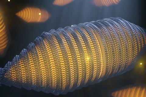
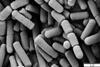
Gas vesicles (GVs) are hollow, cylindrical nanostructures made of a thin protein-based shell and filled with gas. Similar in function to ballast tanks in submarines or fish bladders, many water-based bacteria use these structures to regulate their floatability.
In a paper in Cell, scientists from the Departments of Bionanoscience and Imaging Physics at Delft University of Technology now describe the molecular structure of these vesicles for the first time. These gas vesicles were also recently repurposed as contrast agents for ultrasound imaging.
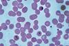
“For example, certain cyanobacteria use gas vesicles to float to the surface in order to harvest light for photosynthesis, a phenomenon sometimes seen at enormous scale in toxic algal blooms,” says Arjen Jakobi, Assistant Professor at the Department of Bionanoscience.
Staying afloat
There are very specific requirements for such structures: for bacteria to stay afloat, GVs must occupy a substantial proportion of the cell, which involves forming compartments that extend over hundreds of nanometres in size.
To maximise floatability, the shell must be constructed from minimal material. At the same time, the shell needs to provide resistance to the pressure from the surrounding water to maintain the ability to float with changes in water depth. GVs have therefore evolved as rigid, thin-walled structures composed of a single protein that repeats many thousands of times to form the GV shell.
“Despite intensive efforts, the molecular structure of GVs and therefore a molecular level understanding of their distinctive properties, has remained elusive”, says Stefan Huber, PhD Candidate in Jakobi’s lab.
“But the very recent development of highly advanced electron microscopy hardware and image processing algorithms has allowed us to solve this structure at almost atomic detail. We can now present the cryogenic electron microscopy (cryo-EM) structure of the GV shell, providing detailed insight into how GVs grow and into the unique evolutionary adaptions that make bacteria float.”
Corrugated wall
The gas vesicle protein GvpA features a corrugated wall structure, typical for force-bearing thin-walled cylinders, similar to the ribbed metal sheet of tin food cans.
Small pores enable gas molecules to move through the shell, while the chemical properties of the gas vesicle’s interior surface effectively repel water. This design allows the GVs to selectively fill with gas. Comparison across a broad range of different bacterial species showed that the elementary design of gas vesicles remained unchanged throughout evolution.
The structure provides fascinating insight into the peculiar molecular features that bacteria have evolved to enable them to float in a watery environment, and it sheds light on the ingenious engineering principles – at the nanoscale – that are required to maintain thin hollow structures under pressure.
Ultrasound potential
The study will also facilitate molecular engineering of gas vesicles for ultrasound imaging.
“In this study we have collaborated with the lab of David Maresca at the Department of Imaging Physics, who approached us to visualise gas vesicles produced in his lab,” says Jakobi. In Maresca’s lab, PhD Candidate Dion Terwiel aims to use gas vesicles contrast agents for ultrasound imaging, by tweaking their genetic code.
The high contrast in density between gas-filled GVs and surrounding cellular structures makes them bright in ultrasound images and their particular properties are a potential improvement over current contrast agents.
“The insights we have gained allow me to re-engineer these acoustic biomolecules more precisely”, says Terwiel. “Since 2014, there has been a renewed interest in gas vesicles as these can serve as the ‘green fluorescent protein’ for ultrasound. Knowing the gas vesicle structure will help us to design acoustic biosensors in order to ‘spy’ on biological processes deep into tissues,” adds Maresca.

No comments yet