It is clear that bacteria and phage coexist in many diverse, complex environments. However, it is perhaps not always clear which party will win when an interaction occurs.
To add to the complexity, recent work has shown that bacteria can tip the balance to survive in the presence of predatory phage when they form biofilms. It is the change in lifestyle that results in protection, not genetics.
Biofilms are structured communities of microorganisms that are attached to a surface and are encased in a self-produced extracellular matrix. The biofilm matrix is dynamic in nature and fulfils multiple functions for the sessile community. This includes nutrient sequestration and water adsorption, shielding the resident cells from environmental stress and competition, and acting as a signalling facilitator for cells both within and outside the biofilm. Research over the last 10–15 years has shown that there is great diversity in the composition of the biofilm matrix across both polymicrobial and between single-species biofilms; however, commonly occurring constituent parts are emerging. These include polysaccharides, extracellular DNA, lipids and proteins, some of which are fibrous in nature. Growing evidence suggests that at least one fibre-forming protein, which provides structural integrity to the biofilm, can additionally provide protection to the resident bacteria from phage predation.
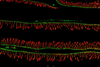
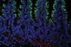
The biofilm matrix of the Escherichia coli biofilm has recently been shown to both sequester and hinder diffusion of a predatory lytic T7 phage. Using a series of bacterial strains, it was shown that the protection offered by the biofilm was dependent on ‘curli’, protein fibres found in the extracellular matrix. Curli fibres made by E. coli were first visualised by transmission electron microscopy and were quickly noted for their high level of insolubility and resistance to proteases. Curli production is dependent on the starvation response of the cells, which can occur in a multi-layered biofilm due to the stratification of cells with respect to a nutrient source. Microscopy analysis has shown that the starved E. coli cells become highly ‘curliated;’ in essence, the protein fibres form a network of ‘cell-moulded baskets’ throughout the intercellular space. It is this protein network that provides structure to the bacterial community, which is the element of the biofilm that is critical to protect the cells from lytic phage. The curli fibres work in two ways. First, they bring the cells in the biofilm together in an intricately connected structure. This physically prevents the phage from entering the biofilm. However, curli can also bind phage, restricting phage mobility by sequestration. The consequence is that the phage are prevented from reaching the cells within the interior of the community. The outcome of this intricate phage–bacterium interaction was revealed through the use of high-resolution microscopy of living biofilms. This allowed the T7 phage and bacterial community to be followed in both space and time. It will now be of interest to see if there are other ways by which the outcome of an interaction between phage and bacteria can be manipulated. The mechanisms known are likely to diversify as the study of bacteria–phage interactions, and the methods by which the analysis is conducted, continues to grow.


No comments yet