Bioleaching bacteria home in on and around grooves and crevices in rare earth element-bearing rocks and form biofilms, potentially offering a route to making the process more efficient, a new study shows.
The research, by a team at Curtin University in Australia, found that colonization and biofilm formation on these REE-containing minerals is not random but selective toward physical imperfections such as grooves and cracks. However, they are not found to be selective towards specific mineralogy or chemical composition on the surface.
The study, ‘Biofilm formation on the surface of monazite and xenotime during bioleaching’ has been accepted for Microbial Biotechnology, an Applied Microbiology International publication.
Close to nil data
Corresponding author Dr Arya van Alin said that to date there has been close to nil data on microbial biofilm formation on the REE-phosphate minerals.
“Conventional extraction metals have severe environmental consequences. The environmental consequences of such methods are worst for REE-containing minerals,” he said.
“Bioleaching offers an eco-friendlier approach - it has been studied for several decades and is used at industrial scale for extraction of copper, gold, nickel and some other metals.
“However, bioleaching of REE-bearing minerals is a fairly new field of study and our understanding of the mechanism behind it is very limited. Simply put, we have been trying to solve a puzzle over time by addressing different questions to better understand the geo-microbial processes behind the bioleaching.
Final pieces of the puzzle
“The current research is one of the last pieces of this puzzle to provide a more detailed understanding of microbial attachment to these minerals, if they form biofilm, whether this attachment is a purely random process or there is some logic behind it, and if the biofilm formation imposes changes on the surface of the minerals.”
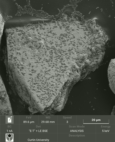
The team investigated monazite, which contains light-REE and xenotime which contains heavy-REE, subjecting them to bioleaching with Klebsiella aerogenes as the microbial agent.
Samples were taken at different time points and studied using a confocal laser scanning microscope (CLSM), scanning electron microscope (SEM) and focused ion beam (FIB)-SEM microscope.
Biofilm formation
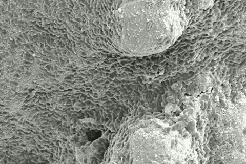
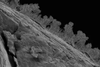
“CLSM and SEM showed that K. aerogenes cells attached to the surface of these minerals and formed a biofilm. Biofilm formation had three stages - an initial attachment of bacteria to the surface within the first 8 hours of exposure, then over time this led to colonisation of more area on the surface (16-24h) and then formation of a mature biofilm (day 2-8),” Dr van Alin said. “However, at some point bacterial cells start detaching from these surfaces which marks the 3rd stage, biofilm dispersion.
“The FIB-SEM analysis revealed a thin-layer structure (average 500 nm thickness) for the biofilm. Compared to the abiotic leaching control (using a sterile acidic media at pH 4), bioleaching resulted in an extensive erosion of the surface.”
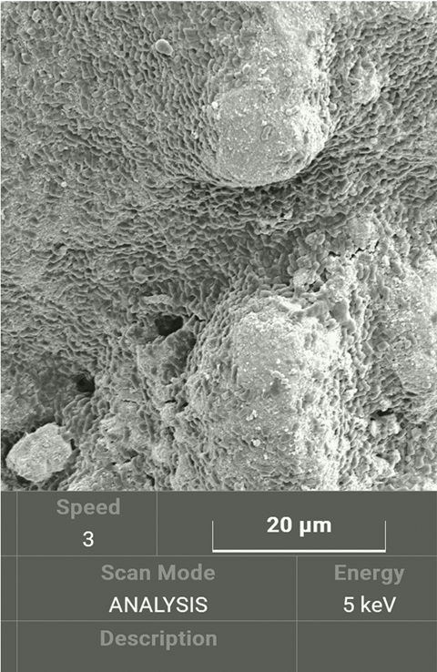
Dr van Alin said the microscopy analysis and 3D modelling of the CLSM-acquired data showed that the attachment and biofilm formation is not purely random.
“Bacterial attachment and biofilm formation was selective on and around the physical imperfections on the mineral surface, sometimes referred to as high-energy sites. These sites give easier access to the nutrient content of the minerals and provide better safety against some of the environmental stress such as the sheer fluid force.
“On the other hand, no selectivity was found toward certain chemical composition or mineralogy.”
Solving the mystery
In recent years, our research group studies on bioleaching of phosphate minerals aimed to solve a puzzle - what are the potential bioleaching models, and the possible mechanism behind these (summarised in a review study by their former PostDoc).
“As mentioned before, this field of research is young, and there are many more gaps to be addressed,” Dr van Alin said.
“In our previous studies, we confirmed the acidolysis as one of the main mechanisms of bioleaching, and proposed complexolysis as another potential mechanism that needs to be confirmed. We also showed there was greater REE dissolution when microbes were attached to the surface of the ores.
Attachment stage
“We also need to better understand the initial attachment stage of the biofilm formation to promote further biofilm formation and as a result enhance the bioleaching efficiency. When we achieve a fundamental understanding of the bioleaching mechanisms, then we can go for a directed engineering and optimisation.”
This study was funded by the Australian Research Council and the Parker CRC for Integrated Hydrometallurgy Solutions and The Institute for Geoscience Research (TIGeR). The project was conducted by AvA. Flowcytometry was designed and developed by AvA and CT. The supervision, parts of the experimental design, and critical review was done by EW (primary supervisor), MC, AK, AP, JE, and HF.
‘Biofilm formation on the surface of monazite and xenotime during bioleaching’ appears in Microbial Biotechnology, an Applied Microbiology International publication.

No comments yet