From microbes in the human gut to symbiotic algae in coral reefs, research in recent decades has increasingly revealed the pivotal roles that microorganisms (or microbial species) play in shaping the biology of host organisms and of broader ecosystems.
For example, some endosymbionts—microbes that live within the cells of a host organism—are known to manipulate the physiology of their hosts to promote their own persistence from generation to generation.
One of the best-known instances of this comes from the insect world, where bacterial endosymbionts in the genus Wolbachia can induce asexual reproduction in their hosts to ensure their transmission to future generations. While this phenomenon was first recognized over 30 years ago, the underlying mechanisms have remained elusive until now.
In a recent study published in Genome Biology and Evolution, titled “Identification of parthenogenesis-inducing effector proteins in Wolbachia”, researchers from the University of Minnesota have identified key Wolbachia proteins that mediate this microbe-induced form of reproduction. The study represents a significant step forward in understanding the molecular mechanisms underlying the manipulation of host reproduction by Wolbachia bacteria, laying the groundwork for unraveling the complex interactions between these endosymbionts and their host organisms.
Prevalent endosymbionts
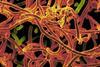
Bacteria in the genus Wolbachia are among the most prevalent endosymbionts on Earth, infecting around half of all insect species. Wolbachia are transmitted from insect mothers to their offspring, and thus male hosts represent an evolutionary dead-end for the bacteria. Because of this, some Wolbachia lineages have evolved the ability to induce parthenogenesis – a form of asexual reproduction that results in all female offspring - in their insect hosts.
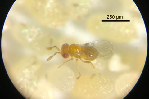
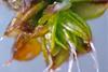
The authors of the new study, Laura Fricke and Amelia Lindsey, focused on Wolbachia strains from two parasitoid wasp host species, Leptopilina clavipes and Trichogramma pretiosum.
These Wolbachia strains are known to induce parthenogenesis in a two-step process: First, the endosymbionts interfere with host chromosome segregation, inducing diploidization, or a doubling of the chromosomes, in unfertilized eggs, giving rise to diploid female offspring. Next, the Wolbachia induce female development in the offspring by interfering with elements of the host’s gene expression system, such as the insect gene transformer, a master regulator that directs the development of individuals towards either male or female phenotypes.
Triggering parthenogenesis
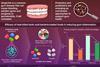
“We hypothesized that the parthenogenesis-inducing Wolbachia strains encoded effector proteins responsible for diploidization and driving female differentiation, key steps in parthenogenesis induction,” explains Fricke. To identify these effector proteins, Fricke and Lindsey looked for genes that were shared by the two parthenogenesis-inducing Wolbachia strains but were absent from all other Wolbachia strains. This led to the identification of two candidate effector proteins, named PifA and PifB (Parthenogenesis inducing factors A and B), that may be responsible for triggering parthenogenesis in host organisms.
Further analysis of PifA and PifB revealed that they exhibit many of the characteristics of eukaryotic host-associated bacterial effector proteins. Structural predictions revealed similarities between PifA and eukaryotic nucleoporins and RNA splicing proteins, while PifB showed structural homology to eukaryotic leucine-rich repeat receptor-like serine/threonine-protein kinases. Intriguingly, PifA also contains a Transformer-like region, hinting that it may be employed by Wolbachia to hijack elements of the insect sex determination pathway. Additional assays demonstrated that both PifA and PifB impact eukaryotic cells and that PifA localizes with atypically dividing nuclei, suggesting a role in manipulating host cell division.
Mystery origin
The discovery of PifA and PifB as candidate effector proteins opens up new avenues for research into Wolbachia-mediated reproductive manipulation. “One interesting question is where these two genes came from,” says Lindsey. “This is a hard question to answer. The surrounding elements in the genome indicate that these genes may have been transferred into the Wolbachia genome by viruses, but we don’t know the original source.” While there are no clear matches to PifA and PifB among insect genomes that have been published, the authors note that this may simply reflect the sampling bias in genetic databases.
The lack of genetic tools for studying Wolbachia and their hosts remains a major challenge to exploring these proteins further. Since standard genetic approaches (e.g., gene knockdown or knockout) are not currently possible, the authors are focusing on a suite of alternatives to help elucidate how these proteins work.
Minuscule size
“From here we are taking a proteomics-based approach to isolate the targets of the Pif proteins in the wasp system,” says Fricke. “We want to see where these proteins localize in the insect embryos during natural infection, and we are currently working to identify which specific insect proteins and RNAs the Pifs interact with,” continues Lindsey.
Even these alternative investigations may be challenging to carry out due to the miniscule size of the parasitoid wasp species they are studying.
“These insects are smaller than a grain of sand, and most people can’t see them without the use of a microscope. It makes many of our experiments quite tricky and often requires a lot of optimization and steady hands to carry out experiments that would be routine in larger insects,” explains Lindsey. Nevertheless, overcoming these obstacles promises to shed new light on the intricate interplay between endosymbiont effectors and the physiology and evolution of their host organisms.


No comments yet