
Study reveals hidden damage in stony corals using 3D imaging and AI
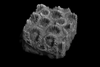
Researchers investigating how disease affects coral structure turned to X-ray microcomputed tomography which generates detailed 3D reconstructions down to microscopic pores, which reveal internal skeletal features, including porosity, thickness and structural orientation, in a non-destructive way.
Registered users, subscribers or members - SIGN IN
To continue reading this article
Membership
Included as a benefit
Members of Applied Microbiology International have unlimited access to The Microbiologist as a benefit of membership. Just sign in with your usual membership credentials.
Find out more about how to join.
Register
FREE
Want to read more before deciding on a subscription? It only takes a minute to sign up for a free account and you’ll get to enjoy more free content each month.
Subscribe
3.75 per month / £35 annually
Get unlimited access to The Microbiologist.