A research team used synchrotron-based X-ray microcomputed tomography (SR-μCT) to non-invasively obtain high-quality 3D images of fresh soybean root nodules, quantifying the volumes of the central infected zone (CIZ) and vascular bundles (VBs).

The study further employed synchrotron X-ray fluorescence imaging to visualize the distribution of iron and zinc within these tissues. This pioneering method enhances the understanding of nodule function in N2-fixation, with potential applications in breeding soybean cultivars for improved nitrogen-fixation efficiency and enhanced root nodule activity.
Legume-rhizobia symbiosis
Nitrogen (N) is crucial for plant growth as it forms essential biomolecules. Modern agriculture relies on synthetic nitrogen fertilizers, which are energy-intensive and environmentally harmful. The legume-rhizobia symbiosis offers a sustainable alternative, efficiently fixing N2 in root nodules. However, the functional significance of nodule tissues in nitrogen fixation is not well understood.
A study (DOI: 10.34133/plantphenomics.0203) published in Plant Phenomics on 29 May 2024 aims to employ advanced imaging techniques to visualize and assess the functional structures in soybean root nodules, enhancing our understanding of nitrogen fixation efficiency.
This study utilized synchrotron radiation micro-computed tomography (SR-μCT) and X-ray fluorescence (SR-XRF) imaging to non-invasively visualize internal structures of fresh soybean root nodules, focusing on central infected zones (CIZ) and vascular bundles (VBs). SR-μCT provided high-quality, high-contrast images without extensive sample preparation, and Biomedisa’s algorithm rapidly segmented nodular tissues.
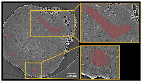
SR-XRF imaging revealed the distinct localization of iron within the CIZ and zinc within the VBs across three soybean genotypes, correlating with nitrogen fixation efficiencies. Despite limitations such as analyzing a single nodule per genotype, this innovative method demonstrated the potential of SR-μCT and SR-XRF for rapid, high-resolution phenotyping, offering valuable insights into nodule structure-function relationships. The study highlighted the utility of these techniques in advancing understanding of plant internal microstructures, suggesting that synchrotron imaging is a powerful tool for future research in this field.
Nitrogen fixation
According to the study’s lead researcher, Leon Kochian: “The proposed methods enable the exploitation of the root nodule’s anatomical features as novel traits in breeding, aiming to enhance N2-fixation through improved root nodule activity.”
In summary, this study highlights the functional importance of CIZ and VBs in soybean root nodules for nitrogen fixation. Using synchrotron-based X-ray microcomputed tomography (SR-μCT), high-quality, non-invasive 3D visualizations and volume quantifications of these tissues were achieved.
Synchrotron X-ray fluorescence imaging further revealed the specific localization of iron and zinc within nodules, showcasing their roles. Future research could leverage deep neural networks for automatic segmentation and synchrotron X-ray fluorescence tomography for detailed 3D mapping, potentially enhancing nitrogen fixation efficiency through advanced soybean breeding strategies.


No comments yet