Research led by a physicist at the University of California, Riverside, shows how viruses form protective shells, or capsids, around their genomes — a process that, while messy and complex, consistently results in highly symmetrical icosahedral structures.
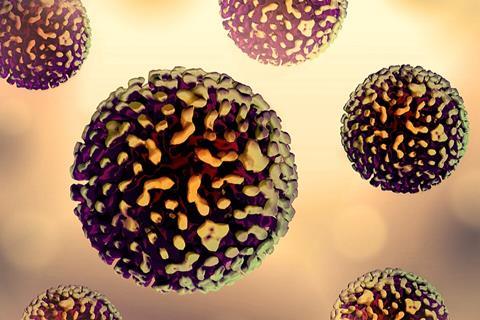
A genome is the complete set of genetic material in an organism. For most organisms, this is DNA, while in some viruses, it is RNA. The genome provides the instructions needed for growth, function, and reproduction. In geometry, an icosahedron is a solid with twenty equilateral triangular faces. Many viruses package their genome inside an icosahedral protein shell, which provides both stability and efficiency in enclosing the genetic material.
READ MORE: Researchers reveal how the SARS-CoV-2 virus acquires its spherical shape
READ MORE: Scientists zoom in on structure of still lethal Ebola virus
“Until now, most studies have focused on simpler systems or relied on artificial constraints,” said Roya Zandi, a professor of physics and astronomy, who led the project. “Our research makes a major leap by simulating, for the first time, the spontaneous formation of larger and biologically relevant capsids, specifically T=3 and T=4 structures, around flexible genomes.”
In viral capsids, T=3 and T=4 refer to the triangulation number, a value that describes how protein subunits are arranged to build a larger, stable, icosahedral structure. A higher number corresponds to a larger, more complex capsid.
Symmetric shells
Zandi and her colleagues used a new simulation framework to capture key biological factors like protein diffusion, genome flexibility, and shape-shifting behavior to model how viral proteins self-assemble into symmetric shells. Their findings, published in Science Advances, could open doors to designing synthetic nanocontainers for medical and biotech uses.
In the following Q&A, Zandi answers a few questions on the research:
Q. What mechanisms drive the virus to form an icosahedral shell despite the “messy and complex” assembly pathway?
A 3,000-nucleotide viral genome, made of RNA, can attract about 180 protein subunits to form a stable shell. Though the process appears chaotic at first, with proteins sticking in the wrong places, their elasticity allows for self-correction, as neighboring forces break faulty bonds. This dynamic leads to the correct, soccer ball-like T=3 icosahedral structure. In the lab, proteins can assemble into irregular shells if no genome is present, or if the genome length is mismatched to the shell. These outcomes highlight the crucial role of the genome and the importance of physical self-correction in achieving the correct icosahedral symmetry.
Q. Why is icosahedral symmetry the preferred structure for viral capsids?
Icosahedral symmetry is the most efficient way to build a strong container from many identical parts. By arranging elastic protein subunits in a perfectly symmetrical way, the virus creates a shell that is both very stable and requires the fewest building blocks. This design gives maximum protection for the genome with minimal cost to the virus. It is nature’s most efficient way to build a shell from many identical proteins.
Q. Is this assembly process conserved across different virus families?
Yes. Most spherical viruses rely on the same physical principle: elastic proteins that can self-correct until they form a symmetrical shell. The details vary between families; for example, larger viruses than T=3 or T=4 often require helper or scaffolding proteins. But the overall strategy of self-assembly into an icosahedral container is universal.
Q. How does the genome influence the structure and symmetry of the viral shell?
The viral genome attracts proteins along its length, forming an initially disordered RNA-protein complex. The genome pulls proteins together, raises their local concentration, and acts as a scaffold to strengthen interactions, aiding shell assembly. Genome size also matters: its radius of gyration influences the most stable shell size. Though capsid proteins can assemble around various RNA lengths or even nanoparticles, our study focused on how a long RNA strand first creates a disordered protein-RNA complex that ultimately forms a stable, symmetrical shell through physical forces.
Q. Experimentally, how did you track the intermediate steps of viral shell formation?
Because viruses are only a few nanometers in size and the intermediate stages are so short-lived, we cannot capture them directly with current experimental tools. Techniques like cryo-electron microscopy and X-ray scattering reveal the final structures, but not the hidden steps in between. That is why simulations were essential. In this study, computer modeling showed the transient intermediates as well as how fragments come together to form a complete shell. This work represents the first published simulations demonstrating how a virus as large as a soccer ball, or an even larger one like hepatitis B (T=4), can assemble step by step, shedding light on stages that experiments cannot see.
Q. How could understanding the assembly process help in designing antiviral drugs or treatments?
By uncovering the short-lived intermediate steps of assembly, we can identify where the process is most vulnerable. Drugs could interfere with these steps by preventing proteins from breaking incorrect bonds, disrupting the elastic corrections they need to self-assemble, or blocking the genome from acting as a scaffold. In each case, the outcome is the same: the virus cannot complete its shell. Without a proper shell, the virus cannot protect its genome and cannot spread infection.
Q. How might your findings help develop synthetic or engineered viruses for gene therapy or nanotechnology?
The same physical rules that viruses use to build their shells can be harnessed for medicine and technology. By tuning the elasticity of proteins and the properties of the cargo inside, researchers could design stable protein shells that package and deliver drugs or genetic therapies safely into cells. This approach also opens the door to creating nanoscale delivery systems for other applications, from targeted medicine to smart materials.
Zandi was joined in the research by Siyu Li, a former graduate student in the Zandi lab, and Guillaume Tresset of Paris-Saclay University in France. Li is now a faculty member in the Physics Department at Cal Poly Pomona.
The research was supported by grants from the National Science Foundation and the University of California Multicampus Research Programs and Initiatives.


No comments yet