Iron storage ‘spheres’ inside the bacterium C. diff — the leading cause of hospital-acquired infections — could offer new targets for antibacterial drugs to combat the pathogen.
A team of Vanderbilt researchers discovered that C. diff (Clostridioides difficile) produces the spheres, called ferrosomes, and that these structures are important for infection in an animal model. The findings, reportedin the journal Nature, are also a rare demonstration of a membrane-bound structure inside a pathogenic bacterium.
Bacteria have long been thought not to contain organelles (such as a nucleus, mitochondria and other specialized structures) like eukaryotic cells, but that biological dogma appears to be incorrect.
Compartmentalizing processes
“The emerging idea that bacteria do compartmentalize biochemical processes in a way similar to eukaryotic cells really flips the field of microbiology on its head,” said Eric Skaar, PhD, MPH, the Ernest W. Goodpasture Professor of Pathology and director of the Vanderbilt Institute for Infection, Immunology, and Inflammation.
Skaar, co-corresponding author Qiangjun Zhou, PhD, assistant professor of Cell and Developmental Biology, and their colleagues were intrigued by findings reported several years ago that some environmental bacteria produce iron-containing ferrosomes.
They knew that the genes in these bacteria were conserved in C. diff and other anaerobic bacteria (bacteria that die in the presence of oxygen), and they set out to determine if C. diff produces ferrosomes to manage its need for iron. Like all living organisms, C. diff requires iron to survive and grow. Skaar and his team have focused on how pathogens like C. diff acquire iron and other metals, with a goal of finding new pathways that could be exploited to ‘starve’ pathogens of essential nutrients.
Limited treatment
C. diff causes about 500,000 infections and more than 29,000 deaths in the United States each year, according to the Centers for Disease Control and Prevention, and treatment options are limited. People taking antibiotics that disrupt the healthy microbes in the gut are at increased risk for C. diff infection, which causes diarrhea and colitis. New strategies for treating this urgent public health threat are needed, Skaar said.
To look for iron inside C. diff, the researchers first drew on expertise and resources in the Vanderbilt Institute of Nanoscale Science and Engineering (VINSE).
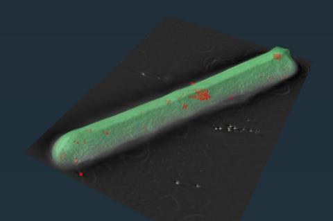
“The best way to look for the accumulation of elements in a small space like a cell is with a method called STEM-EDS, which has not commonly been used for biological samples,” Skaar said. “We were fortunate to have access to a STEM-EDS instrument and collaborators at VINSE, and we quickly proved that there was an accumulation of iron ‘dots’ within the bacterium.”
Co-first authors Hualiang Pi, PhD, and Rong Sun, PhD, led studies to show that those iron dots represented organelles that were important to C. diff infection.
Ferrosome formation
Pi and Skaar’s team found that two genes (fezA and fezB), which are similar to those in environmental bacteria, were required for ferrosome formation. Using C. diff bacteria missing these genes, they showed that ferrosomes are required for C. diff to fully colonize and cause disease in an animal model.
They found that ferrosomes were even more important for C. diff infection in a model of inflammatory bowel disease, demonstrating that these iron-containing structures help the bacterium combat “nutritional immunity” — the host response of producing proteins to bind iron and attempt to starve the pathogen.
Sun and Zhou’s team used cryogenic electron microscopy (cryo-EM) and cryo-tomography to show that the ferrosome structures were encased in a membrane, classifying them as organelles.
Skaar noted that “Vanderbilt’s unique geography” — the proximity of experts in engineering, cell biology and the Medical Center — and specialized tools for STEM-EDS and cryo-EM made the research possible.
Potential targets
The results “establish ferrosome formation and all the factors involved in ferrosome formation as potential targets for new antibacterial drugs against an important infectious disease,” Skaar said.
“Anytime we find new factors involved in host-pathogen interactions and show that they’re important for infection, that opens entirely new opportunities to make classes of antibacterial drugs that have not existed before. That is especially important in the face of rising antimicrobial resistance that we’re seeing globally.” iron
In future studies, the researchers plan to explore how ferrosomes are formed, whether other gut pathogens produce ferrosomes, and whether these structures might be shared in the gut as a source of iron. Skaar is also particularly interested in pursuing the emerging area of bacterial organelles.
“We think our study is a rare demonstration of an organelle in a pathogenic bacterium,” he said. “Now we want to know if there are other subcellular compartments in bacteria that we’re interested in that could teach us about how these cells perform various physiologic processes.”


No comments yet