In a new article published in Nature Communications, researchers from Uppsala Antibiotic Center, Uppsala University and SciLifeLab describe a fundamental mechanism of antibiotic resistance. What happens in a bacterium that is resistant to the antibiotic fusidic acid? With a stop-motion movie at the atomic level, they can show that the resistance protein FusB works almost like a crowbar.
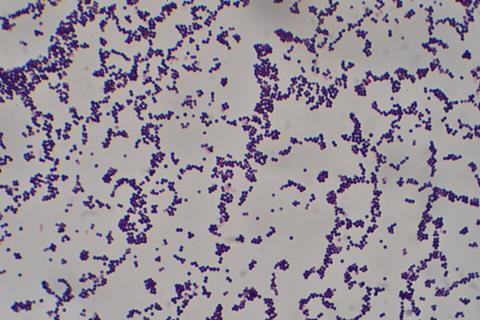
Antibiotic resistance is a global issue that requires action and research at many levels. This study describes the molecular mechanism of the most prevalent type of fusidic acid resistance in the bacterium Staphylococcus aureus.
READ MORE: Scientists discover antibiotic resistance in newly identified bacterium
READ MORE: Scientists discover rules for breaking into Pseudomonas
Antibiotics function in many different ways. Fusidic acid belongs to the large group of antibiotics that target harmful bacteria by blocking their ribosomes, the machineries that translate the genetic code into proteins. However, bacteria can develop resistance to antibiotics in several ways, such as by pumping out a drug or deactivating it. Some resistance mechanisms are more advanced and specific.
Molecular crowbar
“The mechanism behind fusidic acid resistance has been a long-standing mystery,” says Maria Selmer, professor and antibiotic resistance researcher.
In a new article, researchers revealed how FusB, a resistance protein found in clinically resistant strains of the common bacterium Staphylococcus aureus, can rescue ribosomes from fusidic acid. Using electron microscopy, they show that FusB works like a molecular crowbar that frees the blocked ribosome. The researchers describe their method as a “stop-motion movie” at the molecular level.
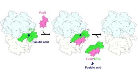
“With traditional methods, we would be too late to the “crime scene”. Thanks to this methodology, we could catch the resistance protein in the act, right as the “crowbar” is “breaking into” the molecular complex,” says Adrián González López, PhD student.


No comments yet