
Drug-resistant bacteria are becoming more common worldwide, posing a major public health challenge. Addressing this problem requires finding new ways to eliminate bacteria, which in turn requires analyzing essential cellular processes that could serve as targets for future antibiotics.
Phages reveal a bacterial weak point
Nature may offer some clues in viruses known as bacteriophages, or “phages”. These viruses infect bacteria and hijack their cellular machinery to replicate. Some of them even inject small proteins that shut down specific bacterial processes, ultimately causing the cell to die.
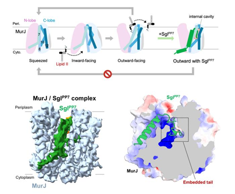
One target is MurJ, a protein that bacteria use to transport lipids needed to build their cell wall. Although several bacteriophage proteins that can inhibit MurJ have been found, their amino acid sequences can be quite varied. How can such different proteins block the same essential target, and do they use the same molecular strategy?
To answer these questions, a research team led by Assistant Professor Hidetaka Kohga of Nara Institute of Science and Technology (NAIST), Japan, examined how a bacteriophage protein called SglPP7 inhibits MurJ. Their study, which was published in PLOS Biology on October 27, 2026, reveals the finer details of how SglPP7 interacts with MurJ and compares its inhibition mechanism with those of other phage proteins that target the same lipid transporter. Co-authors included Mr. Kaito Hosoda, Mr. Sixian Wu, Mr. Hiroyuki Tanaka, Dr. Hideki Shigematsu, Assistant Professor Ryoji Miyazaki, Associate Professor Min Fey Chek, and Professor Tomoya Tsukazaki, all from NAIST.
The researchers used cryo-electron microscopy, a technique that reveals the structures of biological molecules at near-atomic resolution, to determine the structure of the MurJ– SglPP7 complex. They also performed experiments involving targeted protein mutations, bacterial cell lysis, and computer simulations to investigate how the interaction with SglPP7 affects MurJ’s behavior.
Different proteins, same outcome
The results showed that SglPP7 lies along a lipid-binding groove in MurJ and inserts its short C-terminal tail into a deep internal cavity. This interaction stabilizes MurJ in an outward-facing conformation, preventing it from completing the back-and-forth structural changes required to transport lipid. Comparisons with two other known phage proteins, LysM and SglCJ3, revealed that all three stabilize a similar conformation of MurJ despite binding to it in different ways. “These findings suggest convergent evolution toward a common target through diverse inhibitory mechanisms,” says Dr. Kohga.
MICROBIOLOGY NEWS: Register with The Microbiologist for more free articles
Understanding these interactions could help researchers design new drugs and engineer artificial phage proteins with targeted antibacterial activity. “By revealing how phage proteins inhibit MurJ, this study provides a foundation for drug discovery targeting this transporter,” explains Dr. Kohga. “Although still at the basic research stage, these findings could contribute to the development of new antibacterial agents against drug-resistant bacteria.”
Further research efforts in this field will hopefully shed light on antibacterial mechanisms that evolved naturally and bring us closer to a new generation of antibiotics.


No comments yet