Scientists at the Department of Energy’s Oak Ridge National Laboratory have reimagined the capabilities of atomic force microscopy, or AFM, transforming it from a tool for imaging nanoscale features into one that also captures large-scale biological architecture.
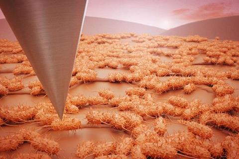
Often called a “touching microscope,” AFM uses a fine probe to feel surfaces at resolutions down to a billionth of a meter. Although powerful, traditional AFM has been limited by its narrow field of view, making it difficult to understand how individual features fit into larger organizational structures.
READ MORE: New microscopy technique reveals dynamic E coli membrane stiffness
READ MORE: High speed atomic force microscopy studies provide insights into influenza A viral replication
Researchers at the Center for Nanophase Materials Sciences, a DOE Office of Science user facility at ORNL, have now overcome this limitation by developing an automated large-area AFM platform. Demonstrated on bacterial biofilms — resilient microbial communities that grow on a range of surfaces — the system connects detailed observations at the level of individual parts with broader views that cover larger areas. This advance offers an unprecedented view of biofilm organization, with innovations for medicine, industry and environmental science.
Broad impact
Biofilms can cause infections, clog pipes, damage equipment and disrupt ecosystems, so they have broad impact. Understanding how biofilms form and organize on surfaces can lead to better infection treatments, more effective cleaning and maintenance strategies, and improved management of natural ecosystems and water quality. Biofilms begin when individual bacteria use sticky proteins or appendages to anchor themselves to a surface. These so-called pioneer cells then grow and recruit others, forming complex, hard-to-remove communities.
“In biofilm research, we’ve often been able to see the trees, but not the forest,” said Ruben Millan-Solsona, a postdoctoral researcher in ORNL’s Functional Atomic Force Microscopy group and the study’s co-leader. “Using the AFM, we could examine individual bacterial cells in detail but not how they organize and interact as communities.”
Liam Collins, an R&D researcher in ORNL’s Functional Atomic Force Microscopy group and the study’s co-leader, added, “This new platform changes that. Now, we can visualize both the intricate structures of single cells and the larger patterns across entire biofilms.”
Key innovation
A key innovation lies in integrating machine learning with the imaging process. “The large-area AFM provides researchers with large-scale, high-resolution views of biofilms,” said Sita Sirisha Madugula, a postdoctoral researcher in ORNL’s Data Nanoanalytics group and co-author of the study. “The integration of machine learning allows us to extract meaningful quantitative data from these massive datasets.”
The team automatically analyzed more than 19,000 individual cells to generate detailed maps of cell properties across extensive surface areas, revealing that bacteria align in honeycomb-like patterns, interconnected by flagella — hair-like appendages that may help with initial attachment and growth.
“Though the biological role of these patterns is still under investigation, they likely play a part in strengthening biofilm cohesion and adaptability,” Collins said.
Nanoscale ridges
To further probe biofilm dynamics, the team tested engineered surfaces with nanoscale ridges — features thousands of times thinner than a human hair. They found that specific patterns could disrupt normal biofilm formation, offering potential strategies for designing antifouling surfaces that resist bacterial buildup.
Published in npj Biofilms and Microbiomes, the research marks a significant leap forward in biofilm science. It is supported by the DOE Office of Science through the Biopreparedness Research Virtual Environment, or BRaVe, initiative, which explores interactions between living systems and materials.
“This collaboration shows what’s possible when scientists from different disciplines come together,” said Scott Retterer, CNMS director and principal investigator for BRaVE. “The advanced tools we’re developing are essential for understanding how organisms interact with materials — a key step to identifying surface properties that resist biofilm formation, which has important applications from healthcare to food safety.”
Power of partnership
The work also benefited from partnerships with the Bredesen Center for Interdisciplinary Research and the University of Tennessee-Oak Ridge Innovation Institute, which contributed to refining the computational algorithms used in the study.
UT-Battelle manages ORNL for DOE’s Office of Science, the single largest supporter of basic research in the physical sciences in the United States. DOE’s Office of Science is working to address some of the most pressing challenges of our time. For more information, visit energy.gov/science.
Topics
- Artificial Intelligence & Machine Learning
- atomic force microscopy
- Bacteria
- Biofilms
- Bredesen Center for Interdisciplinary Research
- Early Career Research
- Economic Equality
- flagella
- Industrial Microbiology
- Innovation News
- Liam Collins
- Marine Science
- Microbiological Methods
- Oak Ridge National Laboratory
- One Health
- Ruben Millan-Solsona
- Scott Retterer
- Sita Sirisha Madugula
- University of Tennessee-Oak Ridge Innovation Institute
- USA & Canada
- Young Innovators

No comments yet