Ebola (EBOV) and Marburg virus (MARV) are highly lethal viruses that cause severe disease in infected patients by extensively damaging the body. This includes the gastrointestinal tract. Severe diarrhea followed by dehydration is a major causes of death in EBOV and MARV disease patients, yet the role of the intestinal lining (epithelium) in these outcomes remain poorly understood.
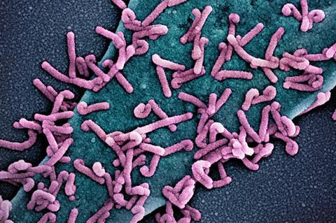
A new study first-authored by Elizabeth Yvonne Flores, PhD, a recent graduate from Boston University Chobanian & Avedisian School of Medicine, BU’s National Emerging Infectious Diseases Laboratories (NEIDL) and the Center for Regenerative Medicine (CReM) of BU and Boston Medical Center, sheds light on the mechanisms behind this damage. The study found that both EBOV and MARV are capable of infecting and replicating within human gut epithelial cells and that the viruses interfere with the cells’ ability to regulate fluid secretion, mirroring the severe symptoms observed in patients.
“This research enhances our understanding of how filovirus infections damage the gut and identifies potential cellular pathways for targeted treatments,” explains co-corresponding author Elke Mühlberger, PhD, professor of virology, immunology & microbiology and an investigator at BU’s NEIDL. “It also highlights how useful iPSC-derived organoids are for studying viral diseases.”
To study actual human gut tissue in a controlled environment, the researchers grew organoids–miniature, 3D structures that mimic human intestinal and colonic tissue from induced pluripotent stem cells (iPSCs), a type of stem cell created in a lab from regular adult cells, like skin or blood cells. They then successfully infected these “mini-guts” with EBOV and MARV, observing that the viruses could replicate within the tissue.
Gene activity signatures
By analyzing gene activity signatures in the infected organoids, they discovered that organoids resembling the small intestine and those resembling the colon responded differently to infection, with more severe dysfunction in the colonic organoids. The infections disrupted key signaling pathways involved in ion and fluid transport in the gut and damaged the structure of the gut lining, including the apical (the outermost surface of a cell) and junctional (the connection between cells) components that control what passes through the intestinal wall.
READ MORE: Human PARP gene could be novel target for viral diseases or immune-mediated disorders
READ MORE: Organoids reveal the secrets of bat immunity
These changes may help explain how these viruses cause the massive fluid loss that leads to life-threatening diarrhea. They also found the infected mini-guts showed a delayed innate immune response, specifically in the production of interferon-stimulated genes, which usually help fight off viruses.
“The organoid platform successfully captures key features of human GI pathology, making it a powerful tool for future research to understand host-pathogen interactions better and identify potential therapeutic targets to treat these deadly diseases,” said co-corresponding author Gustavo Mostoslavsky, MD, PhD, professor of medicine and virology, immunology & microbiology at the school and co-director of CReM.
These findings appear online in the journal PloS Pathogens.


No comments yet