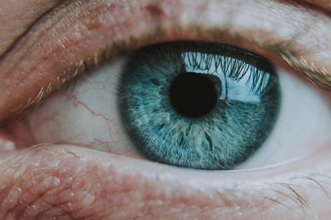
University of Pittsburgh School of Medicine researchers have developed an early-stage, experimental “living eye drop” that uses naturally occurring eye bacteria to support corneal wound healing.
The proof-of‑concept study, published today in Cell Reports, demonstrates that the harmless eye-dwelling microbe Corynebacterium mastitidis can be genetically modified to secrete an anti-inflammatory therapeutic that promotes healing following corneal injury in a mouse model.
“This is the first demonstration that a microbe that lives on the ocular surface could be engineered to deliver a therapeutic that improves eye health,” said senior author Anthony St. Leger, Ph.D., associate professor of ophthalmology and immunology at Pitt and the UPMC Vision Institute. “It opens the door to the idea of ‘living medicine’ for the eye—something you apply once, and it stays, protects and helps the tissue heal.”
Because tears continually wash medications away, treating ocular surface disease often requires multiple daily applications of eye drops. This can limit the effectiveness of therapies for conditions such as corneal abrasions or dry eye disease.
Benign bacterium
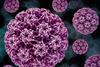
To explore an alternative delivery method, the Pitt team engineered C. mastitidis, a benign bacterium that naturally resides under the eyelid, to continuously secrete cytokine interleukin10 (IL10) – a small protein that regulates inflammation. In mice, corneas that were gently scratched and treated with the engineered bacteria healed faster than those treated with regular bacteria or saline. When the IL10 receptor was blocked, this benefit disappeared—confirming the therapeutic effect was IL10-dependent.
The researchers also created a version of the microbe that releases human IL10, which improved wound closure in lab-grown cells that make up the outermost layer of human cornea and reduced inflammatory signaling in human immune cells. These studies offer an initial indication that the approach could eventually be adapted for use in people, though substantial development remains.
“What makes this exciting is that the system is modular,” St. Leger explained. “We built it so you can swap in different genes—different cytokines, growth factors or other proteins—to tailor the therapy to specific eye diseases.”
Developing off-switches
Though promising, the technology is still in early development. The researchers note that many steps must be completed before any clinical translation is possible, including developing built‑in “off switches,” a way to safely and reliably remove or deactivate the engineered bacteria after they are no longer needed.
READ MORE: Common bacteria discovered in the eye linked to cognitive decline
Conditions such as severe dry eye, inflammatory disorders of the ocular surface and traumatic corneal injuries affect millions of Americans each year. While this study does not yet establish a clinical therapy, it provides a foundation for exploring whether engineered live biotherapeutics could one day offer more sustained delivery of anti-inflammatory or regenerative molecules to the eye.
“In my lab, we don’t typically build tools from the ground up,” St. Leger added. “Seeing a measurable improvement in healing in an animal model using something we engineered was incredibly rewarding, and it points us toward intriguing possibilities for future research.”
The research was led by Jackie Shane, Ph.D., with additional contributions from Matthew Evans, Yannis Rigas and Robert Shanks, Ph.D., all of Pitt.


No comments yet