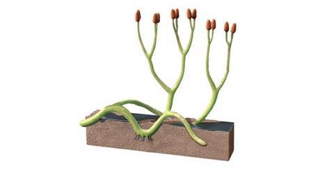
Researchers from the Natural History Museum and Sainsbury Laboratory Cambridge University (SLCU) have identified a new species of ancient symbiotic fungus preserved within a 407-million-year-old plant fossil from Scotland. The discovery provides unprecedented three-dimensional insight into one of the earliest known plant–fungus partnerships, known as mycorrhiza.
Beyond this discovery, the advanced microscopy techniques used to distinguish the fungus from the surrounding plant cells open a powerful new way to identify fossilised life forms. By analysing their unique light signatures — a kind of natural fingerprint preserved through time — scientists can detect traces of organisms long after their DNA has vanished.
READ MORE: Symbiotic and pathogenic fungi may use similar tools to manipulate plants
READ MORE: Fungi paved the way for life on land hundreds of millions of years earlier than previously thought
Published in New Phytologist, the paper describes a new species of arbuscular mycorrhizal fungus, Rugososporomyces lavoisierae, forming a symbiotic relationship with the early land plant, Aglaophyton majus — the second fungal species known to have been hosted by this plant.
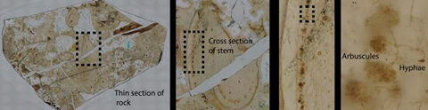
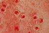
The fossil from the Windyfield Chert, Scotland provides the most detailed evidence to date that early land plants engaged in complex symbiotic relationships with multiple fungal species over 400 million years ago. The fossil, held at the National Museum of Scotland, Edinburgh, was prepared and studied by the Natural History Museum’s scientific associate Dr Christine Strullu-Derrien, who co-led the study.
Rare mycorrhizas
This fossilised relationship was found to closely resemble modern arbuscular mycorrhizal associations that continue to play a vital role in plant nutrition and soil health today.
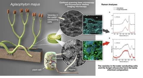
“Mycorrhizas are very rare in the fossil record and have never been found in the Windyfield Chert before,” said Dr Christine Strullu-Derrien. “The presence of the arbuscule shows that the fungus wasn’t parasitising on the plant or feeding on it after death – instead, there was a symbiotic association. The fungus would have provided minerals like phosphorus in return for sugars from the plant in a way that benefits them both.”
Dr Paul Kenrick, a fossil plant expert at the Museum, and co-author of the study, added: “It’s extraordinary to find such ancient evidence of a symbiotic relationship. It appears that symbioses were a necessary part of allowing plants to adapt to life on land; this symbiosis resembles that observed in modern liverworts or hornworts; these plants like the early land plants don’t have roots. Finding out more about how these relationships developed can contribute to our knowledge of the past, present and future.”
Light signatures
The interdisciplinary team brought together specialists from the Natural History Museum who found the new fungus and conducted brightfield microscopy and confocal microscopy with the Muséum d’Histoire Naturelle in Paris; the Sainsbury Laboratory Microscopy Core Facility, who conducted confocal, fluorescence lifetime imaging microscopy (FLIM) and Raman imaging; and the Cambridge Graphene Centre, responsible for Raman spectroscopy.
The combined use of advanced imaging and spectroscopy applied for the first time to a fossil plant enabled the team to distinguish fossilised fungal and plant tissues based on their unique light signatures, marking a breakthrough that could transform how scientists’ study ancient life in the future.
“By combining confocal fluorescence lifetime imaging with Raman spectroscopy, we can chemically identify ancient microscopic life forms with remarkable precision. Our new technique is opening an exciting new window on life’s earliest chapters,” said Dr Raymond Wightman, Manager of the Sainsbury Laboratory Microscopy Core Facility who led the FLIM imaging work.
Powerful tool
The implications of this discovery extend far beyond the immediate findings.
“This is just the start,” said Professor Schornack who co-led the study. “By applying these methods to the fossilised remains of different organisms, we now have a powerful new tool to tell apart structures that may look similar but differ in their fine ultrastructure, for example ancient arthropods, plants and fungi.”
“This technique adds a new dimension to how we identify, describe and discriminate fossilised ancient life, using the unique light signals these materials emit as a kind of fingerprint. Although the original biological material is carbonised and no DNA remains, these optical signatures preserve vital clues to their identity.”
Using these techniques with other fossils from the Windfield and nearby Rhynie cherts, researchers aim to understand how early symbioses evolved and how plants and fungi first learned to coexist.
Christine Strullu-Derrien, Raymond Wightman, Liam McDonnell, Gareth Evans, Frédéric Fercoq, Paul Kenrick, Andrea Ferrari and Sebastian Schornack (2025) An arbuscular mycorrhiza from the 407-million-year-old Windyfield chert identified through advanced fluorescence and Raman imaging. New Phytologist. DOI: https://doi.org/10.1111/nph.70655
Topics
- Aglaophyton majus
- Christine Strullu-Derrien
- Ecology & Evolution
- fossil record
- Fungi
- Healthy Land
- Microbiological Methods
- mycorrhiza
- Natural History Museum
- Paul Kenrick
- Raymond Wightman
- Research News
- Rugososporomyces lavoisierae
- Sainsbury Laboratory Cambridge University
- Sebastian Schornack
- Soil & Plant Science
- symbiosis
- UK & Rest of Europe

No comments yet