What if people could detect cancer and other diseases with the same speed and ease of a pregnancy test or blood glucose meter? Researchers at the Carl R. Woese Institute for Genomic Biology are a step closer to realizing this goal by integrating machine learning-based analysis into point-of-care biosensing technologies.
The new method, dubbed LOCA-PRAM, was reported in the journal Biosensors and Bioelectronics and improves the accessibility of biomarker detection by eliminating the need for technical experts to perform the image analysis.
Traditional medical diagnostic techniques require doctors to send patients’ blood or tissue samples to clinical laboratories where expert scientists carry out the testing procedures and data analysis.
READ MORE: Nanorobot hand made of DNA grabs viruses for diagnostics and blocks cell entry
READ MORE: Good as gold - improving infectious disease testing with gold nanoparticles
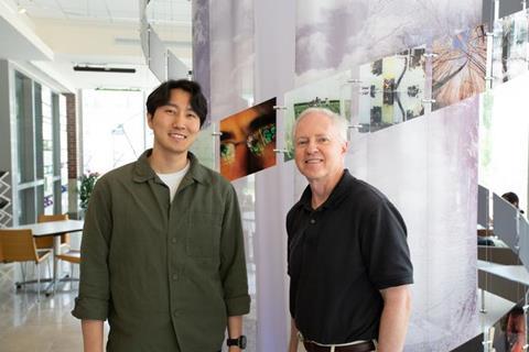
“Current technologies require patients to visit hospitals to get diagnostics which takes time. A lot of people also have barriers where more appointments may not be financially or spatially feasible,” said Han Lee, first author of the study and a graduate student in the Nanosensors research group. “I think that we can make a difference by developing more point-of-care technologies that are available for people.”
Point-of-care diagnostics
Point-of-care diagnostics are performed and yield results at the site of patient care, whether at home, the doctor’s office, or anywhere in between. This allows for lower cost, easy-to-use, and rapid tests that can help inform next steps. Some examples already adopted in everyday life include urine pregnancy tests, COVID-19 antigen testing kits, and blood glucose meters which allow people with diabetes to respond to dips and spikes in their blood glucose levels throughout the day.
In the point-of-care field, researchers are investigating new ways to integrate these types of technologies into patient care settings, such as appointments with specialists like oncologists or oral surgeons. This would help to reduce the time and financial burden on patients, while improving real-time decision making for physicians.
Bacterial infection
“Physicians say they would like something similar to when you go in with bacterial infection. They do a test on you right there and then send you home from your appointment with the right antibiotics that will treat the particular bacteria that you have,” said Brian Cunningham (CGD leader), a professor of electrical and computer engineering. “So why not do a similar thing for choosing the right anti-cancer drug or determining if the drug you’ve been taking for a couple weeks is starting to work or not.”
Previously, the group reported a new biosensing method called Photonic Resonator Absorption Microscopy, or PRAM, to detect molecular biomarkers—molecules in the body whose presence and levels indicate healthy or disease states. PRAM enables the detection of single biomarker molecules including nucleic acids, antigens, and antibodies; common biosensing techniques instead detect the cumulative signal of hundreds to thousands of molecules.
Going for gold
Cunningham said, “Basically, what we’re doing is shining a red LED light at the bottom of a sensor. Then on the top of the sensor, molecules are landing and getting detected whenever they have a tiny particle made out of gold—which we call gold nanoparticles or AuNPs—attached to it.”
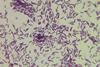
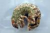
The images generated using PRAM depict a red background with little black spots sprinkled across it. But while these images themselves seem relatively simple, obtaining an accurate count requires a trained eye that can decipher what spots truly correspond to the AuNP-tagged biomarker molecules.
“There are many kinds of artifacts such as dust particles or aggregates of the nanoparticles. If you don’t have a lot of experience, it’s hard to distinguish them,” Lee said. “The conventional counting algorithm that we’ve been using requires adjusting a lot of parameters to get rid of those artifacts.”
Machine learning
In order to move this process out of the laboratory and make it better suited for point-of-care environments, Lee proposed integrating machine learning into the image analysis process.
“Han really on his own developed an interest in machine learning after taking a class here at the university just to learn about it,” Cunningham said. “He came to me one day and said that he thought he could make a machine learning algorithm count our black spots more accurately.”
Compared to other biosensing techniques, PRAM lends itself well for incorporating deep learning algorithms because it generates microscope images, rather than just detecting optical signals. But because these algorithms are only as good as the data that trained it, Lee decided to image the same samples using both PRAM and scanning electron microscopy.
Tiny black holes
The AuNPs, which are 1000 times smaller than human hair and only show up as small black spots in the PRAM images, can be more clearly visualized on the electron microscope. In a time intensive process, Lee cross referenced every spot in the PRAM images with the electron microscope images to obtain highly accurate data for the machine learning training set.
“Finding the right spot to compare to was actually very challenging because it’s like finding a needle in a desert. One way that I devised was to create a reference point, like a lighthouse in a sea. Then from there we can find the exact same spot for registrations,” Lee said.
The resulting deep learning-based method, called Localization with Context Awareness, integrated with PRAM, enables real-time, high precision detection of molecular biomarkers without needing the eyes and experience of a technical expert. When tested, the team found that LOCA-PRAM surpassed conventional techniques in accuracy, detecting lower levels of the biomarkers and minimizing rates of false-positive and negatives.
Impactful changes
“The whole journey of my PhD was started because I wanted to make changes in the point-of-care field,” Lee said. “I just want to do everything in my power to develop more advanced technologies that can be impactful in the future.”
The publication, “Physically grounded deep learning-enabled gold nanoparticle localization and quantification in photonic resonator absorption microscopy for digital resolution molecular diagnostics” can be found at https://doi.org/10.1016/j.bios.2025.117455 and was supported by the National Institutes of Health, USDA AFRI Nanotechnology grant, and National Science Foundation.


No comments yet