
New research from the Royal Veterinary College (RVC) has found that antibodies against Johne’s disease can be detected in the blood of newborn calves after they have received colostrum from their mothers. The findings suggest these antibody measurements could provide a new way to determine when calves are first exposed to the disease. This will support future efforts to better understand how Johne’s disease spreads and ultimately improve disease control and cattle welfare.
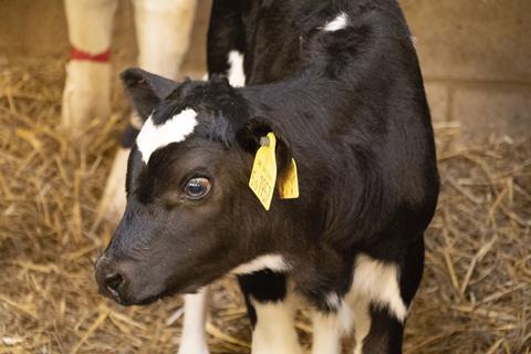
Johne’s disease is a chronic and incurable infectious disease of cattle caused by Mycobacterium avium subspecies paratuberculosis (MAP). Cattle are thought to become infected during the first few weeks or months of life, but signs of disease often do not develop for several years. If infected, the disease progressively damages the lining of a cow’s small intestine, which causes chronic inflammation and can lead to severe weight loss, low milk yields, diarrhoea and death.
MICROBIOLOGY NEWS: Register with The Microbiologist for more free articles
The disease causes major animal welfare concerns and economic losses worldwide, with more than half of UK dairy herds estimated to be affected. Current surveillance programmes mainly focus on identifying infection in adult cattle, leaving important questions about exposure during early life.
Blood samples
Seeking to better understand early life exposure, researchers from the RVC analysed blood samples from 38 newborn dairy calves and their dams on a commercial dairy farm in south-west England. These samples had originally been collected as part of routine monitoring of whether calves had successfully absorbed antibodies from colostrum.
The research team then measured MAP-specific antibodies in the remaining (residual) serum samples and used statistical modelling to examine how these antibody levels related to the mother’s Johne’s disease status and the efficiency of antibody transfer from colostrum.
The study found that calves born to cows with MAP antibodies had higher levels of MAP-specific antibodies than calves born to cows without detectable antibodies. The relationship between antibody levels and the amount of antibody absorbed from colostrum also differed depending on whether the mother was MAP-seropositive or MAP-seronegative. Together, these findings are consistent with MAP-specific antibodies being passed from cow to calf through colostrum.
While these antibody measurements are not a diagnostic test for infection in newborn calves, they may eventually complement existing herd surveillance and risk assessment strategies that could help improve disease management and animal welfare.
Straightforward test
Jonathan Hedgecock, RVC veterinary student graduate and lead author of the study, said: “Continued research into new testing strategies for Johne’s disease is important with more work still needed. Many calves are already blood sampled for serum total protein at a young age, so being able to test calves at this age would be straightforward as we wouldn’t need to take additional blood samples nor would we have to wait until they develop disease as adults.”
MICROBIOLOGY ON TAP: Get full access to all The Microbiologist articles from just £2.17 a month
Dr Steven Van Winden, Associate Professor of Population Medicine at the RVC, and supervisor of this research, said: “Johne’s disease is unusual because infection typically occurs very early in life, yet we often cannot detect its consequences until years later. Our study shows that antibodies against the disease can be transferred from cow to calf and measured in newborn calves. While much more work is needed, this opens up an intriguing possibility for studying exposure risk during one of the most important periods in a calf’s life.”




No comments yet