
Viruses are sneaky intruders, hijacking the internal machinery of host cells and replicating out of sight. Understanding these dynamics is the first step to developing effective antiviral strategies that could stop infections, but scientists have largely been left in the dark because there are limited tools to study how viruses move in space and time inside live infected cells, according to Joyce Jose, associate professor of biochemistry and molecular biology at Penn State.
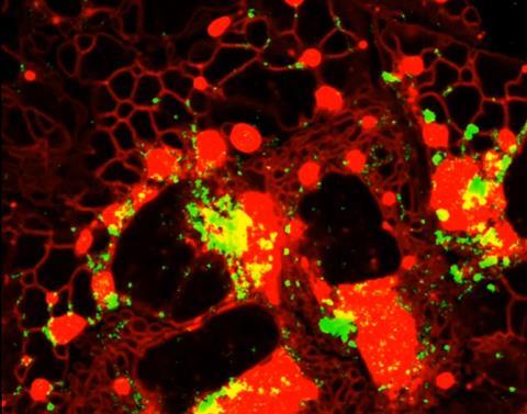
Now, a team of researchers from Penn State led by Jose have filmed the genetic material of Zika and chikungunya viruses — two types of RNA viruses that can cause severe disease and even death in humans — moving inside live infected cells in the laboratory. The research team developed a fluorescent molecular tag that’s built into the virus’s own genetic material, allowing them to watch in high resolution and in real time how the viral RNA genetic material interacts with its host and replicates. They then used the same tool to identify host proteins that are crucial to life cycles of the viruses.
The novelty of the technique, called a dual-action aptamer tag and described in the study published in the journal Nature Communications, is the tag’s two-for-one role — tracking the genetic material and the interacting host proteins, according to the research team. In addition, since the tag doesn’t affect the virus’s life cycle, scientists can now study how viral genetic material engages with its host during its native infectious life cycle. Jose said this approach could be applied to study other RNA viruses like dengue, hepatitis C and rhinovirus.
“Live imaging like what we’ve accomplished here is a very powerful tool and is something we’ve been working toward for the last eight to nine years,” said Jose, senior author on the study. “Existing tools like electron microscopy capture fixed images but you lose the temporal resolution, the moment-to-moment interactions between virus and host inside the cell. It’s the difference between a photograph and a video.”
Deadly families
Zika is a member of the Flaviviridae family, which includes West Nile, dengue and yellow fever viruses, and is typically transmitted by vectors — living organisms like mosquitoes that carry the pathogen and infect another organism. Chikungunya, which is also a mosquito-borne virus, is a member of the Togaviridae family, like Eastern equine encephalitis virus.
In recent years, there have been a number of outbreaks of Zika and chikungunya infections around the world, explained Jose, but there are currently no good antiviral treatments for either infection. In order to develop new therapeutics, scientists need to understand the virus-host cell interaction — the who, what, where and when of how the virus exploits the host cell — so that they can identify potential targets to interrupt the chain of action and stop the virus from spreading.
However, imaging viral RNA, especially during an active virus infection, is difficult, Jose explained. It’s hard to manipulate the viral genome in a way that doesn’t affect the virus’s replication cycle because the genome is so compact and the material is all essential for the virus’s survival. Scientists might witness one replication cycle but then the virus dies.
Mango solution
Here, the research team used an aptamer tag called Mango-II. These short, single-strand DNA or RNA sequences are inserted directly into the genome. Typically, the tag is inserted into the regulatory region of the genome but doing so in viral RNA would interfere with the virus’s ability to replicate. Instead, the researchers inserted the tag into the protein-coding region of live Zika and chikungunya RNA, which hadn’t been done before, to visualize viral RNA.
When the tagged viruses infected commercially available human cells in the lab, the researchers could observe the Zika and chikungunya viruses as they actively translated and packaged viral genomes in live host cells and where these activities occurred under high-resolution advanced microscopy. The Mango-II tag forms secondary structures on the single-stranded viral RNA and binds to a stain that emits a bright green fluorescent color under the microscope. When the virus undergoes its double-stranded RNA replication stage, the fluorescent color goes dark.
The two viral families displayed distinct movement patterns. Zika’s viral RNA is anchored to the endoplasmic reticulum, the main manufacturing and transportation network inside a cell, whereas chikungunya’s viral RNA travels more widely and rapidly along the plasma membrane. This live, real-time imaging validated what has been previously published in the research literature, the research team explained.
Double action
But the Mango-II tag wasn’t just a fluorescent marker. It also allowed the researchers to extract the tagged viral RNA from the infected cells and the host proteins that interacted with the viral genome, providing insight into which proteins from the host cell the viruses depend on to survive. They analyzed the material and after comparing their results against two controls, they identified with high confidence more than 200 host proteins that interacted with Zika virus and a smaller subset for chikungunya virus, with 132 common host factors shared between the two viruses.
MICROBIOLOGY NEWS: Register with The Microbiologist for more free articles
Among the shared host factors, they found that a protein called SFPQ was vital to the life cycle of both Zika and chikungunya viruses. When the researchers depleted SFPQ in the host cell using a technique called short hairpin RNA (shRNA), which genetically dials down the protein’s expression in the cells, viral production of both viruses dropped drastically.
“Despite being very different viruses, we can say that it’s a host protein that’s important for both viruses to replicate,” Jose said. “And when we looked at it under the live imaging again, we could see it.”
Benjamin Kropff, a doctoral student in Jose’s lab, also contributed to the paper. A full list of other authors can be found in the paper.
Funding from the National Institutes of Health’s National Institute of Allergy and Infectious Diseases under awards 1R01AI176505, 1R01AI168169, 1R21AI178533 and 1R01AI177352, and United States Department of Agriculture for the NACA 58-3022-2-038 contributed to this work. This content is the sole responsibility of the authors and does not necessarily reflect the views of the funders.






No comments yet