A new study outlines how an innovative tool can be used to help uncover the reasons why phages succeed or fail when used to target bacterial infections.

This study, led by Ruizhe (Erez) Li as a PhD student in the Bakshi Lab, Engineering Department was presented in a talk at the Viruses of Microbes (VOM) event earlier this year and was awarded the Sustainable Microbiology journal award for best scientific presentation at the conference.
Ruizhe described how the University of Cambridge team had developed a high-throughput single-cell phage infection tracking assay that allows them to quantify the kinetics of each step of phage infection in individual bacterial cells and correlate these parameters with the host’s physiological state.
This breakthrough provides a systems-level understanding of phage efficacy, revealing when and why phages succeed or fail in clearing their targets.
“Moreover, by understanding how infection parameters depend on bacterial physiology, we can design combination therapies that modulate bacterial physiology, making bacteria more susceptible to phage-mediated eradication, paving the way for more targeted and reliable antimicrobial strategies,” he said.
Lytic phage infections
Ruizhe is a PhD student at the Bakshi Lab (https://www.bakshilab.net/), which focuses on analysing the factors that cause the emergence and spread of antibiotic resistance, and developing and testing alternative treatment strategies to combat resistant infections. “In our group, we are addressing the challenge of harnessing lytic phages as effective therapeutics to treat antibiotic resistant infections by understanding how host cell physiology influences phage infection kinetics,” Ruizhe said.
The research from the Bakshi lab has recently shown how cell physiology affects antibiotic persistence and resistance in bacteria (eg. Caño Muñiz et al, Manuse et al).
“Like antibiotics, phages also depend on the physiological state of their bacterial hosts, and an incomplete understanding of these dependencies can lead to tolerance and phenotypic resistance, making treatments unpredictable and less effective. Our goal is to determine how host physiology and its variability impact phage efficacy, enabling us to quantify escape and resistance rates at the single-cell level. By integrating these measurements into predictive models, we aim to improve the reliability of phage-based interventions at the population level.”
Combining expertise
The assay to track lytic phage infections in individual cells was developed through a collaboration between the Bakshi Lab in the Engineering Department and Dr. Diana Fusco’s lab in the Physics Department, combining expertise in single-cell imaging, microfluidics, and machine-learning with genetic engineering of phages to advance the understanding of phage-bacteria interactions.
To study lytic phage infections at the single-cell resolution and with sufficient throughput, the researchers developed a microfluidic-based infection assay that allows a large number of independent host cell lineages to be maintained under controlled homogeneous growth conditions, while they are infected by phages.
They had to design and optimise the microfluidic channels in this device to ensure that phages can diffuse efficiently to isolated linear colonies maintained in each trench (led by Charlie Wedd, a PhD student in Bakshi Lab from the EPSRC Sensor CDT program at Cambridge).
To track the infection steps, they used time-resolved fluorescence and brightfield imaging to monitor steps of infection from T7 phages that were engineered to contain a genomically integrated fluorescent marker (genetically engineered by Dr. Temur Yunusov, a postdoc in the Fusco lab).).

This data was processed using machine-learning-based analysis tools (originally developed by a former PhD student in the Bakshi lab, Georgeos Hardo (currently an Assistant Professor · United Arab Emirates University) and adapted to this task by Charlie Wedd and Ruizhe) to precisely track and quantify each stage of infection within individual cells, from adsorption to lysis.
Pipeline of information
This single-cell phage infection tracking pipeline provided unprecedented information about the dynamics of each step in the infection process and its relation to the host cell parameters, such as their size, ribosome content, growth rate, etc.
READ MORE: The Microbiologist special issue on Bacteriophages
The next step was to harness the physiological heterogeneity of the infected cells and connect it to the heterogeneity of the infection kinetics, by building a mathematical model to relate the associated variabilities. This model of viral gene expression was built based on basic assumptions of how phage gene transcription was dependent on the viral RNAP, while the translation kinetics is dependent on the abundance of host ribosomes and the corresponding viral mRNA. This model was fit to the experimental data and the fit predicted that as the viral DNA replication progresses exponentially and the mRNAs are rapidly produced off the DNA, host ribosome occupation rapidly saturates.
Limiting factor
This result suggests that the host’s translation capacity becomes a limiting factor in phage gene expression, and possibly determines how many new phage particles can be made in a cell - an observation that was later verified by Ruizhe using phage infections on a ribosome-labelled bacterial strain, where the translation capacity of each cell could be directly estimated from the fluorescence signals.
This analysis led to one unexpected finding, as Ruizhe explained: “Previous bulk studies which compare averages of population-level data from experiments conducted under different growth conditions or with different phage or host mutants have concluded that phage production scales linearly with the infection-to-lysis time.
“However, when we looked into the heterogeneity of infection kinetics within a population (from a pure host-phage genotype and under control constant growth condition), we found that instead the host physiological parameter (here ribosome content) was a stronger predictor of the phage gene expression and the time of infection to lysis had a weaker predictive power.
“This finding highlights the clarity of mechanistic interpretation from data collected at single-cell resolution, which does not blur and average the dynamics of unsynchronised infections across physiologically heterogeneous infected cells, as typically done in bulk measurements.” [noted by Ruizhe’s supervisor Dr. Bakshi].
Predicting phage effectiveness
Dr. Bakshi’s group at the Engineering Department is using this approach for developing physiology-aware treatment strategies to antibiotic resistant bacterial infections. By uncovering how bacterial physiology influences phage infection dynamics, we can predict conditions under which phages are most effective and identify scenarios where resistance or escape is likely, Ruizhe said.
“This knowledge is crucial for designing robust phage-based treatments that minimize failure rates. Furthermore, by leveraging small-molecule or antibiotic interventions to manipulate bacterial physiology, we can enhance phage efficacy, making treatments more predictable and adaptable to clinical and industrial applications.
“Furthermore, our platform provides a powerful tool for screening both engineered and natural phages, enabling precise characterization of their infection dynamics and a deeper mechanistic understanding of their efficacy, ultimately guiding the development of optimized phage therapies.”
Additionally, Ruizhe and his advisor Dr Bakshi are currently working towards developing a rapid phage susceptibility testing platform leveraging their microfluidic-microscopic phage infection tracking assay, for quickly screening and identifying potent engineered/natural phages to eradicate a target pathogenic bacterium.
Dr Somenath Bakshi (Department of Engineering, University of Cambridge) has recently been awarded an ERC Consolidator Grant to study how bacterial physiology shapes phage infection dynamics, with the aim of improving the reliability of phage-based strategies in clinical settings.
Next steps
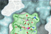
This study so far focuses on model phages. But phages are being used and developed to treat AMR infections such as the staph phage in head picture to treat MRSA infections. The Bakshi group is planning to work with such clinically relevant phages and their target in the next step.
“We also need to refine predictive models by integrating large-scale single-cell data with population-level infection dynamics.
“Finally, validating these insights in physiologically relevant infection models will be crucial for translating our findings into real-world therapeutic and biocontrol applications.”
The research has not yet been published but is based on the following two papers:
Topics
- Applied Microbiology International
- biocontrol agents
- Charlie Wedd
- Clinical & Diagnostics
- Community
- Diana Fusco
- Georgeos Hardo
- Innovation News
- Microbiological Methods
- One Health
- Phage Therapy
- Ruizhe (Erez) Li
- Temur Yunusov
- UK & Rest of Europe
- United Arab Emirates University
- University of Cambridge
- Viruses
- Viruses of Microbes


No comments yet