As summer kicks into full gear and people are spending more time outside, there’s one thing on many people’s minds — ticks. Tick season is starting earlier and lasting longer, and ticks are popping up in areas they haven’t been found before, expanding the risk of tick-borne viruses.
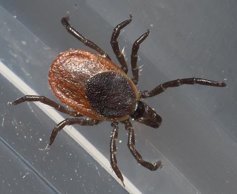
One emerging tick-borne virus in North America is the Powassan virus (POWV), which can cause encephalitis, seizures, paralysis and coma. Rates of POWV infections have increased in recent years and currently, there are no treatments available, according to Joyce Jose, associate professor of biochemistry and molecular biology at Penn State.
READ MORE: Tunnel-building virus: How Zika transmits from mother to fetus
READ MORE: New testing approach improves detection of rare but emerging Powassan virus spread by deer ticks
“We don’t know much about the structure of this virus, but we need to know the structure in order to come up with strategies to treat and prevent infection,” Joyce said, explaining that her team, which includes researchers from Penn State, the University of Minnesota and the US Department of Agriculture, has built a high resolution, 3D structure of POWV. They published their findings today (July 9) in the journal Science Advances.
Deadly family
POWV is a member of the Flaviviridae family, which includes West Nile, dengue and yellow fever viruses. It’s transmitted by vectors, which are living organisms that carry the virus and infect other organisms. In this case, the vectors are ticks.
Because POWV can cause serious health problems, it’s been challenging for researchers to study it in its natural form, Joyce said. Typically, they inactivate the virus by modifying it with chemicals or ultraviolet light. However, these processes often damage the virus, making it difficult to determine its structure at a high resolution, she explained.
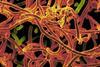
Instead, the team used a surrogate to study POWV. Using the yellow fever vaccine virus, a weakened strain of the yellow fever virus that’s less infectious, they swapped out two protein genes and replaced them with two genes that encode the structural proteins found on the surface of POWV. These proteins — envelope proteins and membrane proteins — are arranged on the surface of POWV in a herringbone-like pattern. It’s a standard and safe practice that has been used to study the surface structure of other types of viruses, Joyce said.
Imaging the virus
They then imaged the virus in Penn State’s Cryo-Electron Microscopy (cryo-EM) facility. Cryo-EM is a technique that allows researchers to determine the 3D structure of proteins and viruses at near atomic resolution. With cryo-EM, researchers can see every molecule in the virus, enabling the team to capture every angle and reconstruct it into a 3D structure featuring the details of the surface proteins.
“When I started my research, viruses used to look like blobs because the resolution was so low,” Joyce said. “Now, we know how every molecule sits on the surface, as well as which ones are more exposed and accessible.”
Understanding the structure of the virus is necessary for understanding viral transmission, something Joyce said is not well understood. Interestingly, she noted, the team found that the type of host the virus transmits through isn’t determined by the structural proteins on the surface of the virus but by the virus’s nonstructural proteins.
“One thing we learned is that the viruses that are transmitted by mosquitoes cannot be transmitted by ticks and vice versa, but we don’t understand what prevents them from cross-transmitting,” she said. “Knowing what the virus looks like — what proteins are on the surface — is the first step. It can shed light on virus-host and virus-vector interactions and how to prevent them.”
Visual details
Vaccines and treatments typically target surface proteins, as well, so this revealing these visual details could potentially inform future therapeutics, Joyce said. Next, the team plans to continue to examine the factors that influence how viruses are transmitted.
Other Penn State authors on the paper include Ibrahim Moustafa, Sung Hyun Cho and Anqi Wang. First author Sayan Das earned a graduate degree at Penn State and senior author Susan Hafenstein was at Penn State at the time of the research; they are now at the University of Minnesota. Dana Mitzel of the U.S. Department of Agriculture also contributed to the paper. A full list of contributors is available here.
Funding from the National Institutes of Health and the U.S. Department of Agriculture supported this work.


No comments yet