
Previous studies have shown that Klebsiella pneumoniae can alter lung metabolism and change how macrophages, immune cells that help clear bacteria, respond to infection. In severe empyema, antibiotics and drainage are central to care, yet inflammation can persist and damage the barrier that normally keeps fluid from leaking into the lung’s air sacs.
Much of the earlier research examined either the bacterium or the immune response at a single point in time. That left an open question about how inflammation changes during illness and whether blocking multiple signaling pathways could reduce the resulting tissue injury.
A study (DOI: 10.48130/targetome-0026-0055) published in Targetome on 05 November 2026 by Xudong Wu’s team, Nanjing University, reports that combined treatment with curcumin and evodiamine reduced signaling through both enzymes and eased lung injury in a mouse model.
Pleural fluid
The researchers profiled small molecules and measured gene activity in pleural fluid collected over three weeks from one patient with severe K. pneumoniae empyema. The chemical profiles pointed to changes in taurine metabolism, while the gene activity data identified three genes at the center of an inflammation-associated network, MAPK8, JAK2 and TP53.
When the team mapped these genes onto publicly available single-cell data from severe lung disease, it found elevated expression in macrophages and in type II alveolar cells, which help maintain the air sacs. Computer simulations that modeled switching off individual genes linked the network to inflammatory signaling and impaired barrier function.
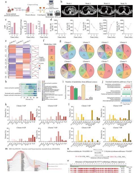
The team then searched drug-response databases for compounds predicted to reverse the patient’s gene activity patterns. Molecular simulations pointed to curcumin and evodiamine, and tests in cultured human lung cells found that curcumin preferentially stabilized MAPK8, while evodiamine modestly stabilized both MAPK8 and JAK2.
Testing in animals
To test the compounds in animals, the researchers began treatment before exposing mice to lipopolysaccharide, a component of the bacterium’s outer membrane. They measured lung fluid accumulation, protein leakage into the airways, tissue damage and inflammatory markers. Both compounds reduced lung swelling and injury when given individually.
Together, they reduced activation of both signaling pathways and brought two circulating inflammatory markers, interleukin-1 beta and tumor necrosis factor alpha, close to levels seen in control mice. Lung tissue from mice given the combination also showed fewer infiltrating macrophages and neutrophils. A separate experiment pairing inhibitors of the same pathways likewise reduced tissue damage, supporting the choice of MAPK8 and JAK2 as targets for further study.
MICROBIOLOGY NEWS: Register with The Microbiologist for more free articles
The study links molecular changes observed during a patient’s illness to two pathways that can be targeted to reduce acute lung injury in mice. The clinical data came from one patient, and the animal experiments tested a short-term inflammatory response to a bacterial component. Larger patient studies and animal studies involving live infection could clarify whether the approach can limit lung damage while preserving bacterial clearance. Further work will also need to establish how the compounds are absorbed and distributed, assess their safety and determine whether they can serve as starting points for drugs used alongside antibiotics.



No comments yet