With the global population aging rapidly, millions of joint replacement surgeries are performed each year. However, this common procedure carries the risk of a devastating complication: periprosthetic joint infection (PJI).
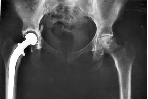
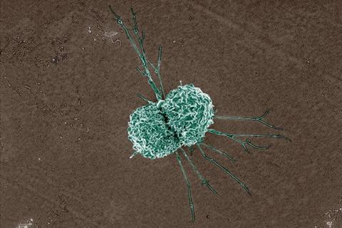
When an implant becomes infected, bacteria like Staphylococcus aureus trigger massive host cell death. The immune system’s failure to promptly clear these dead cells creates a toxic environment that sustains inflammation, hinders tissue repair, and allows the infection to persist, often leading to surgical failure.
While regular exercise is known to generally improve immune function, the exact molecular mechanisms bridging physical activity and localized immune defense in deep tissues like bones and joints have remained elusive. A breakthrough study published in the Journal of Sport and Health Science by researchers from Renji Hospital, Shanghai Jiao Tong University School of Medicine, has finally decoded this “molecular language.”
Molecular mechanisms
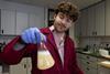
The research team, led by Dr. Bing Yue and Dr. Xinhua Qu, established a direct mechanistic link between regular treadmill exercise and enhanced host defense against PJI in murine models. Through comprehensive bulk RNA sequencing and proteomics, they identified “musclin”—a myokine secreted by skeletal muscles during contraction—as the critical messenger.
“We found that injecting mice with musclin replicated the protective effects of regular exercise,” said Dr. Zhiwei Fu, co-first author of the study. “Musclin significantly reduced the bacterial load and mitigated inflammatory bone loss. It doesn’t act as a traditional antibiotic to kill bacteria directly, but rather as a potent ‘damage-control’ signal that resets the local immune environment.”
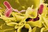
The core mechanism uncovered by the team centers on a process called “efferocytosis”—the ability of macrophages to act as cellular scavengers and engulf dead, apoptotic cells. In a PJI microenvironment, this crucial function is severely impaired. The researchers discovered that circulating musclin binds directly to the formyl peptide receptor 2 (FPR2) on the surface of macrophages.
This specific ligand–receptor interaction triggers a profound metabolic reprogramming within the macrophages. It shifts the cells away from pathological, excessive glycolysis and restores functional mitochondrial oxidative phosphorylation (OXPHOS). This metabolic switch provides the necessary energy and signaling cues to drastically enhance the macrophages’ efferocytosis capacity. By efficiently clearing the dead cellular debris, musclin breaks the vicious cycle of inflammation.
Therapeutic potential
The study demonstrated substantial therapeutic potential when translating these findings into a treatment model. When musclin was administered alongside a conventional antibiotic (oxacillin), the combination therapy synergistically reduced bone destruction, suppressed tissue fibrosis, and successfully restored the adaptive immune response, outperforming antibiotic treatment alone.
This research highlights the musclin–FPR2–efferocytosis axis as a novel therapeutic target.
“Our findings provide scientific support for developing ‘exercise-mimetic’ host-directed immunotherapies,” added the research team. “For patients in the perioperative or rehabilitation phases of joint replacement, safely mimicking the physiological signals of exercise could offer a powerful new adjunctive strategy to combat challenging implant-associated infections.”
Topics
- apoptosis
- Asia & Oceania
- Bacteria
- Bing Yue
- Disease Treatment & Prevention
- efferocytosis
- Immunology
- Infection Prevention & Control
- Infectious Disease
- joint replacement surgery
- macrophages
- Medical Microbiology
- Microbial Genetics
- One Health
- periprosthetic joint infections
- proteomics
- Proteomics & Enzymology
- Public Health
- Renji Hospital
- Research News
- RNA sequencing
- Shanghai Jiao Tong University School of Medicine
- skeletal muscle
- Staphylococcus aureus
- Xinhua Qu

.jpg)
No comments yet