
Alejandro Fernández Llorente works as a predoctoral researcher at Centro de Biología Molecular Severo Ochoa (CBM-CSIC), a prestigious biomedical research centre located in Madrid, Spain.
My passion for microbiology, and specifically virology, began halfway through my university degree while I was studying Biochemistry. I discovered the existence of a vast microscopic world, how it functions, its implications, and how we can ‘ally’ ourselves with these microbes to fight many other diseases together.
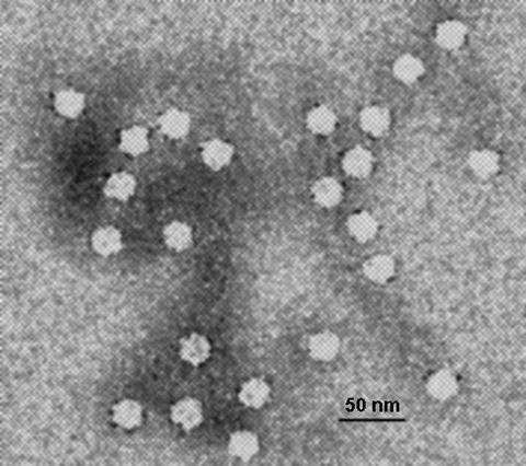
This sparked in me a desire to work in medical virology once I had finished my studies, and undertaking a Master’s degree in Microbiology opened the doors to a laboratory where I worked with a giant virus: poxvirus. Poxviruses are the microbes that fascinate me most. They have a curious microscopic barrel-like shape, are large in size and possess a high degree of autonomy when parasitising cells.
But what surprises me most about poxviruses is that they propel themselves over long distances thanks to actin comets—extensions induced by the viruses in the parasitised cell to push and catapult them. From there, I moved on to working with a tiny virus, one of the smallest known: the parvovirus. Despite its size, this virus harbours enormous complexity, which I am trying to unravel in my current research.
Monday morning: time to get up early and welcome the new week. As soon as I arrive at my research centre, I remember that millions of microscopic creatures are waiting for me behind the doors of the cell culture laboratory. These tiny friends are cells that I seeded onto a Petri dish a few days ago.
Thanks to the nutrients I provided to keep them well-fed throughout the weekend, these cells have reproduced and multiplied (‘germinated’, as if they were seeds). Looking at the dish under the microscope, I can see that they have taken up almost all the space. They are ready to work, just like me.
So, with great care and tenderness, I lift them from their plastic ‘bed’ to wake them up. I seed some of them onto various plates for the experiment I’ll be carrying out this week, while I transfer others to a new Petri dish so that the cells can continue reproducing until Friday and remain active for future experiments.
Relatively relaxed
Tuesday, the relatively relaxed day of the week. While carrying out other experiments, I set aside a few minutes of my day to observe the cells I seeded the day before under the microscope. They look good. They landed on the plastic of the plates and spread their entire cell bodies as if they were fried eggs: the cell nucleus remains firm like the yolk whilst the cytoplasm spreads out like the white. Tomorrow the cells will be ready to make their debut in the experiment!
Natural disaster
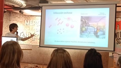
Wednesday, the time has come for infection. After two days in the new plates, the cells have resumed their life cycle without any issues and are multiplying normally. This is essential for them to be infected properly by our second protagonist: the minute virus of mice, abbreviated as MVM.
MVM is a rodent parvovirus that successfully infects cells (from mice and beyond, including human cells too) that reproduce uncontrollably, such as tumour cells and the cells I work with. As this virus is harmless to healthy cells, the laboratory has been studying it for many years for its potential use against cancer.
Without further ado, I shatter the harmony in which the cells are living. Like a natural disaster caused by a higher power, aided by a vacuum, I remove all the nutrients on which they were living comfortably. And suddenly, a liquid floods the cells. A liquid laced with parvovirus.
In a matter of seconds, a storm of viral particles descends upon the cells, which they will infect without mercy. When the storm subsides, I remove the liquid by aspiration and return the nutrients to the cells so that it appears as though nothing untoward has occurred, allowing them to resume their cell cycle as normal. However, not everything is as it was before. MVM is inside the cells now.
Tools of the trade
Thursday: it’s time to collect samples. After a whole night of infection, the MVM parvovirus has caused changes in the cells that are now ready for analysis. I will study these changes for the rest of the week in search of any findings that will allow me to advance my research, and to do so I use molecular biology techniques.
There are several techniques I use in the laboratory to study the infection, procedures that have been optimised thanks to the efforts and experience of researchers generation after generation. One of these techniques, the one I use most, is immunofluorescence. With this technique, I stain specific proteins in the sample I have collected with a fluorescent dye and, by looking at it under the microscope, I can locate them and check whether they are outside the cells, inside them, and even in which cellular compartment.
Another technique (the one I like best) is the Western blot. It requires a great deal of precision, two days’ work, and I can ruin the experiment at numerous critical moments if I am not careful. That’s why every time I get a good result from a Western blot, it’s cause for celebration.
Protein mixture
For this technique, I need to dissolve the cells from the samples and everything associated with them—such as the parvoviruses infecting them—to create a protein mixture.
The Western blot separates all the proteins from this complex mixture and specifically detects the one I’m interested in.
Finally, a technique I use less frequently is the polymerase chain reaction, better known as PCR. Thanks to PCR, I can analyse the MVM parvovirus genome for interesting mutations or detect changes in the expression of cellular and viral genes, amongst other applications.
All these methodologies allow me to study MVM infection from different angles to better understand how it works and to target it for cancer therapy.
Every result matters
Friday, data analysis day. Once I have successfully collected the results from the molecular techniques I developed, it is essential to analyse them rigorously.
Every finding is interesting; every high-quality result matters, even if it differs from what was expected. After all, Nature is what it is, not what one wants it to be.
Before the day draws to a close and gives way to the weekend break, Monday’s Petri dish has filled with cells requiring my attention. To keep them active, I seed some of the cells in another dish with enough nutrients for them to survive the whole weekend and, by the following Monday, be ready for a new experiment.
Alejandro Fernández Llorente is a pre-doctoral researcher at Universidad Autónoma de Madrid. He works at Centro de Biología Molecular Severo Ochoa (CBM-CSIC), a centre of excellence in biomedical research, to understand the molecular mechanisms by which the MVM parvovirus enters the cells it infects. This microbiological research aims to therapeutically direct the virus’ infection towards human cancer cells so that it infects and kills only the diseased cells, leaving the healthy ones intact.


No comments yet