Dr. Peijun Zhang, Director of the Electron Bio-Imaging Centre (eBIC) at the UK’s national synchrotron facility Diamond Light Source, reveals how Cryo-ET is powering some of the most important advances in vaccine research.
Over the last 50 years, vaccines have saved at least 154 million lives – that’s six lives a minute, every day, for five decades. We must recognise that vaccines are one of humanity’s greatest scientific and humanitarian achievements and that behind every vaccine is a story of groundbreaking science.
One of the biggest game-changers in making vaccine development faster and more effective have been advances in imaging techniques. These tools have allowed us, scientists, to visualise and see whether a vaccine is achieving the desired mechanism of action – a crucial step for validating new candidates before they ever reach a syringe.
Vaccines work by imitating the infection generated by a pathogen, a disease-causing organism, to engage the body’s natural defences. They contain either weakened or inactivated parts of the pathogen (known as antigens) or, in the case of newer vaccines, the genetic blueprint (DNA or RNA) for producing those antigens. Regardless of the form they take, vaccines aim to teach the immune system to recognise a specific pathogen without exposing the body to the full risks of disease.
Immune response
When exposed to an antigen, the immune system generates a response by creating antibodies, proteins that specifically bind to that antigen. These antibodies neutralise the pathogen or flag it for destruction by other immune cells. Crucially, this initial immune response also leads to the creation of memory cells, allowing the body to create a rapid, stronger defence if it encounters the pathogen again in the future. Advances in imaging techniques have enabled scientists to capture exactly how this happens.
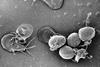
In recent years, Cryogenic Electron Tomography (Cryo-ET) has allowed researchers to capture and observe viruses and cellular processes in the most native cell environments with near-atomic resolution.
Cryo-ET consists of flash-freezing biological samples to capture high-resolution, two-dimensional projection images from a series of viewing angles using electron microscopy that are then used to reconstruct the structures in three-dimensional volumes. It provides unparalleled insights into molecular structures through the imaging of single particles. When used at the cellular level, it also reveals interactions inside cells with unprecedented detail.
Key advances
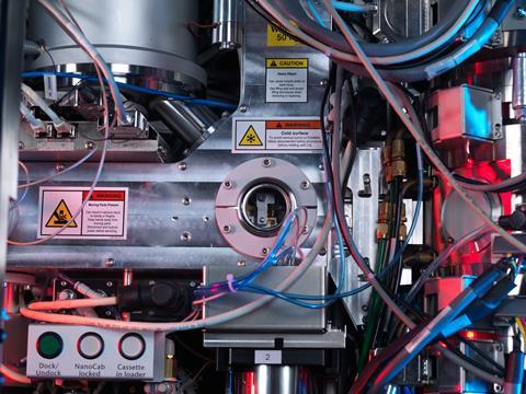
Within the Harwell Science and Innovation Campus in Oxfordshire, the UK’s national synchrotron facility, Diamond Light Source, is powering some of the most important advances in vaccine research. Working as a giant microscope, Diamond uses intense beams of X-rays, electrons, and infrared light to probe the structure of matter at the atomic and molecular levels.
Within Diamond, the eBIC (Electron Bio-Imaging Centre) provides scientists with state-of-the-art experimental equipment and expertise in the field of cryo-electron microscopy, for both single particle analysis and cryo-tomography.
During the early stages of the COVID-19 pandemic, for example, Cryo-ET studies conducted using Diamond’s facilities enabled researchers to rapidly verify the structure of the SARS-CoV-2 spike protein, a crucial antigen used in nearly all COVID-19 vaccine designs. One of the critical insights gained through this high-resolution imaging was the importance of presenting the spike protein in its pre-fusion conformation.
Viral surface proteins
Key viral surface proteins, like the spike, undergo significant structural changes as they prepare to fuse with human cells. Vaccines can present and stabilize these proteins in their pre-fusion form and stimulate the immune system to produce antibodies that are significantly more potent at neutralising the virus. These pre-fusion antibodies bind to vulnerable regions exposed only before fusion, blocking infection before it can begin.
In contrast, post-fusion forms of the protein are structurally altered and less accessible, often resulting in weaker and less protective immune responses. By enabling scientists to capture and stabilise the spike protein in its most immunogenic form, Diamond’s imaging capabilities directly contributed to vaccine candidates, such as Oxford-AstraZeneca’s, that could generate stronger, broader, and more durable protection.
Infectious disease threats
Beyond COVID-19, Diamond’s capabilities have been pivotal in uncovering how viruses like HIV-1 infiltrate host cells and continue to be utilised with other human and non-human pathogens like Polio or Foot-and-mouth disease virus (FMDV), a highly contagious viral disease of livestock.
As infectious disease threats evolve, so too must our tools. Diamond researchers are already utilising machine learning and artificial intelligence to select regions of interest and capture the right images or to visualise the three-dimensional confirmation of the images’ proteins.
eBIC and the wider Diamond Light Source stand at the frontier of this effort through collaborations with synchrotrons across the world helping to further enable the kind of precise, real-time structural analysis that will be essential for rapidly responding to future outbreaks. With the convergence of advanced imaging, data science, and biological insight, facilities like Diamond are not only supporting vaccine development but also accelerating it.
Dr. Peijun Zhang is Director of the Electron Bio-Imaging Centre (eBIC) at Diamond Light Source and Professor of Structural Biology at the University of Oxford.
After completing a Ph.D. in Molecular Biophysics at the University of Virginia and postdoctoral research at the National Cancer Institute, she joined the University of Pittsburgh, where she was promoted to Associate Professor.
Her research integrates cryo-electron microscopy, biochemistry, and computation to study complex molecular assemblies and cellular machinery. As Director of eBIC, she leads one of the world’s foremost cryo-EM centres, advancing structural biology and training the next generation of imaging scientists.
Topics
- Be inspired
- COVID-19
- Cryogenic Electron Tomography
- Diamond Light Source
- Electron Bio-Imaging Centre
- Emerging Threats & Epidemiology
- Foot-and-mouth disease virus
- Harwell Science and Innovation Campus
- HIV-1
- Immunology
- Infectious Disease
- Microbiological Methods
- One Health
- Oxford-AstraZeneca
- Peijun Zhang
- polio
- SARS-CoV-2
- UK & Rest of Europe
- University of Oxford
- University of Pittsburgh
- University of Virginia
- Vaccinology
- Viruses


No comments yet