
Influenza A virus (IAV) causes up to 650,000 annual respiratory deaths worldwide and can trigger severe pneumonia, ARDS, and persistent pulmonary fibrosis.
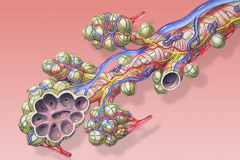
The alveoli, which are the lung’s gas-exchange units, are the primary site of IAV damage, lined by two key cell types: alveolar type 1 (AT1) cells and alveolar type 2 (AT2) cells. How these cells respond differently to IAV has long been unclear.
Mouse models and cell culture studies
To address this gap, a collaborative team led by Professors Bin Cao and Ivan Fan-Ngai Hung conducted a comprehensive investigation, published in the Chinese Medical Journal, combining in vivo mouse models and novel primary cell culture systems. First, the team performed time-resolved immunofluorescence staining on lung tissues from mice infected with the pandemic A/California/07/2009 (H1N1) strain.
As early as 3 days post-infection (dpi), IAV antigens were detected almost exclusively in AT2 cells, with minimal viral presence in AT1 cells. By 7 dpi, AT2 cell coverage in the alveolar epithelium dropped sharply, confirming heightened in vivo vulnerability of AT2 cells to IAV.
To further investigate the mechanism underlying this difference, the researchers established an in vitro primary alveolar epithelial cell culture model and an influenza virus infection system. This model successfully reproduced the infection pattern observed in vivo, confirming that AT2 cells supported higher levels of viral replication than AT1 cells.
Antiviral immune activation
Interestingly, the greater susceptibility of AT2 cells was not explained by differences in viral entry receptor abundance. Transcriptomic analysis revealed a key difference in antiviral immune activation. Although AT2 cells contained higher levels of influenza viral gene segments, they showed weaker expression of antiviral response genes compared with AT1 cells.
In particular, genes related to type I and type III interferon responses, interferon regulatory pathways, interferon-stimulated genes, cytokines, and chemokines were induced more rapidly and strongly in AT1 cells. In AT2 cells, however, these protective immune responses were delayed and attenuated. The researchers further validated these findings using quantitative PCR.
MICROBIOLOGY NEWS: Register with The Microbiologist for more free articles
The findings overturn the assumption that AT1 cells (with their larger surface area) are IAV’s primary initial target and distinguish IAV infection from non-infectious injuries (e.g., hyperoxia, which mainly damages AT1 cells).
The new primary alveolar cell models also provide a platform for future pathophysiology research and antiviral drug screening. The team notes further validation in human cells and with diverse IAV strains (including highly pathogenic avian influenza) is needed to confirm clinical relevance.
Topics
- alveoli
- antiviral immune systems
- ARDS
- Asia & Oceania
- Bin Cao
- cell culture
- Clinical & Diagnostics
- Disease Pathology
- Disease Treatment & Prevention
- Immunology
- Infection Prevention & Control
- Infectious Disease
- influenza A virus
- Interferons
- Ivan Fan-Ngai Hung
- Medical Microbiology
- mouse model
- One Health
- pneumonia
- pulmonary fibrosis
- Research News
- Virology
- Viruses



No comments yet