
Herpes simplex virus type 1 (HSV-1), the virus best known for causing cold sores, infects a large portion of the world’s population and can remain dormant in nerve cells for decades. In recent years, researchers have found evidence linking HSV-1 to neurological conditions such as Alzheimer’s disease, but scientists still do not fully understand how the virus affects the brain.
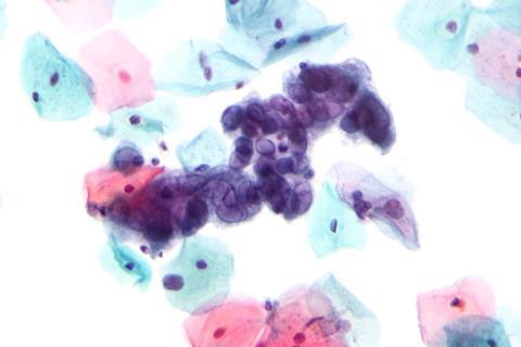
A recent study published in Neurophotonics sheds light on that question by showing how HSV-1 changes the way human neurons produce and use energy.
Lab grown brain tissue
Using a laboratory-grown model of human brain tissue, researchers discovered that HSV-1 rapidly rewires cellular metabolism. Infected neurons initially boost their energy production, but over time this heightened activity is accompanied by signs of stress and impaired mitochondrial function.
According to author Maria Savvidou, a senior researcher in the Department of Biomedical Engineering at Tufts University, “Our findings provide new insight into how persistent viral infection may reshape neuronal metabolism and potentially contribute to processes associated with neurological disease.”
The researchers grew human neurons in three-dimensional scaffolds that mimic some of the structure of brain tissue. This allowed them to study infection in a setting that more closely resembles the human brain than traditional cell cultures grown on flat surfaces. The tissues were exposed to low levels of HSV-1 and monitored for 10 days.
Optical imaging
To track what was happening inside the cells, the team used advanced optical imaging techniques that rely on the natural light emitted by molecules involved in energy production. This enabled repeated measurements of the same living tissue over time, allowing the researchers to capture the dynamic metabolic response to infection rather than relying on single end-point measurements.
MICROBIOLOGY NEWS: Register with The Microbiologist for more free articles
The virus spread through the tissue over time, but the infected neurons remained largely alive and intact throughout the experiment. This was an important finding because it showed that substantial metabolic changes can occur even before extensive cell damage becomes apparent.
Changes in metabolism
One of the most striking changes involved lactate, a molecule produced when cells break down glucose for energy. Lactate is often thought of as a metabolic byproduct, but the study suggests it may play a more active role. Shortly after infection, neurons released increased amounts of lactate. Later, neurons reused some of this lactate as an alternative fuel source, helping meet the increased energy demands of infection.
The imaging data showed that infected neurons entered an unusually active metabolic state within a day of infection. They increased activity in both of the cell’s main energy-producing pathways, indicating that the virus places substantial demands on the cell’s resources.
As the infection continued, signs of cellular stress became more apparent. By days 7 and 10, the neurons showed evidence of oxidative stress, a condition in which harmful reactive molecules can accumulate and damage cellular components. The researchers also detected increasing levels of lipofuscin, an autofluorescent pigment that tends to build up in aging or stressed cells.
The study suggests that lactate may help neurons adapt to the demands of viral infection by serving as an alternative fuel source. However, this adaptation may come at a cost. Sustained reliance on this altered metabolic state was associated with growing oxidative stress and other signs of cellular dysfunction.
Changes to the mitochondria
The cells’ mitochondria, often described as the cell’s power plants, also showed signs of altered function. While the neurons continued producing energy, the data suggested that the process was becoming less balanced and potentially less efficient over time.
When the researchers combined multiple measurements of cellular metabolism, they found that infected and uninfected tissues became increasingly different as the experiment progressed. The largest differences appeared after 10 days of infection, indicating that HSV-1 causes a gradual but substantial shift in neuronal metabolism.
According to the researchers, the work demonstrates the value of noninvasive imaging for studying how viruses affect living brain tissue. By revealing metabolic changes that appear before major cell damage occurs, the approach could help scientists identify early warning signs of neurological disease and better understand the connection between persistent viral infections and brain health.
Topics
- Alzheimer’s disease
- brain
- Clinical & Diagnostics
- Disease Pathology
- Disease Treatment & Prevention
- herpes simplex 1
- Immunology
- Infection Prevention & Control
- Infectious Disease
- lactate
- Maria Savvidou
- Medical Microbiology
- neurons
- One Health
- optical microscopy
- Pharmaceutical Microbiology
- Research News
- USA & Canada
- Virology
- Viruses






No comments yet